Magnesium »
PDB 5grf-5h2f »
5gue »
Magnesium in PDB 5gue: Crystal Structure of COTB2 (Ggspp/MG2+-Bound Form) From Streptomyces Melanosporofaciens
Enzymatic activity of Crystal Structure of COTB2 (Ggspp/MG2+-Bound Form) From Streptomyces Melanosporofaciens
All present enzymatic activity of Crystal Structure of COTB2 (Ggspp/MG2+-Bound Form) From Streptomyces Melanosporofaciens:
4.2.3.146;
4.2.3.146;
Protein crystallography data
The structure of Crystal Structure of COTB2 (Ggspp/MG2+-Bound Form) From Streptomyces Melanosporofaciens, PDB code: 5gue
was solved by
T.Tomita,
S.-Y.Kim,
T.Ozaki,
A.Yoshida,
T.Kuzuyama,
M.Nishiyama,
with X-Ray Crystallography technique. A brief refinement statistics is given in the table below:
| Resolution Low / High (Å) | 34.15 / 1.80 |
| Space group | P 1 21 1 |
| Cell size a, b, c (Å), α, β, γ (°) | 58.698, 100.373, 108.937, 90.00, 90.00, 90.00 |
| R / Rfree (%) | 19.1 / 23.1 |
Magnesium Binding Sites:
The binding sites of Magnesium atom in the Crystal Structure of COTB2 (Ggspp/MG2+-Bound Form) From Streptomyces Melanosporofaciens
(pdb code 5gue). This binding sites where shown within
5.0 Angstroms radius around Magnesium atom.
In total 4 binding sites of Magnesium where determined in the Crystal Structure of COTB2 (Ggspp/MG2+-Bound Form) From Streptomyces Melanosporofaciens, PDB code: 5gue:
Jump to Magnesium binding site number: 1; 2; 3; 4;
In total 4 binding sites of Magnesium where determined in the Crystal Structure of COTB2 (Ggspp/MG2+-Bound Form) From Streptomyces Melanosporofaciens, PDB code: 5gue:
Jump to Magnesium binding site number: 1; 2; 3; 4;
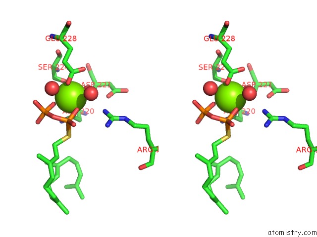
Magnesium binding site 1 out of 4 in 5gue
Go back to
Magnesium binding site 1 out
of 4 in the Crystal Structure of COTB2 (Ggspp/MG2+-Bound Form) From Streptomyces Melanosporofaciens

Mono view

Stereo pair view
Mono view
Stereo pair view
A full contact list of Magnesium with other atoms in the Mg binding
site number 1 of Crystal Structure of COTB2 (Ggspp/MG2+-Bound Form) From Streptomyces Melanosporofaciens within 5.0Å range:
|
Magnesium binding site 2 out of 4 in 5gue
Go back to
Magnesium binding site 2 out
of 4 in the Crystal Structure of COTB2 (Ggspp/MG2+-Bound Form) From Streptomyces Melanosporofaciens

Mono view

Stereo pair view
Mono view
Stereo pair view
A full contact list of Magnesium with other atoms in the Mg binding
site number 2 of Crystal Structure of COTB2 (Ggspp/MG2+-Bound Form) From Streptomyces Melanosporofaciens within 5.0Å range:
|
Magnesium binding site 3 out of 4 in 5gue
Go back to
Magnesium binding site 3 out
of 4 in the Crystal Structure of COTB2 (Ggspp/MG2+-Bound Form) From Streptomyces Melanosporofaciens

Mono view
Stereo pair view
Mono view
Stereo pair view
A full contact list of Magnesium with other atoms in the Mg binding
site number 3 of Crystal Structure of COTB2 (Ggspp/MG2+-Bound Form) From Streptomyces Melanosporofaciens within 5.0Å range:
|
Magnesium binding site 4 out of 4 in 5gue
Go back to
Magnesium binding site 4 out
of 4 in the Crystal Structure of COTB2 (Ggspp/MG2+-Bound Form) From Streptomyces Melanosporofaciens

Mono view

Stereo pair view
Mono view
Stereo pair view
A full contact list of Magnesium with other atoms in the Mg binding
site number 4 of Crystal Structure of COTB2 (Ggspp/MG2+-Bound Form) From Streptomyces Melanosporofaciens within 5.0Å range:
|
Reference:
T.Tomita,
S.-Y.Kim,
K.Teramoto,
A.Meguro,
T.Ozaki,
A.Yoshida,
Y.Motoyoshi,
N.Mori,
K.Ishigami,
H.Watanabe,
M.Nishiyama,
T.Kuzuyama.
Structural Insights Into the COTB2-Catalyzed Cyclization of Geranylgeranyl Diphosphate to the Diterpene Cyclooctat-9-En-7-Ol Acs Chem. Biol. V. 12 1621 2017.
ISSN: ESSN 1554-8937
PubMed: 28463490
DOI: 10.1021/ACSCHEMBIO.7B00154
Page generated: Sun Sep 29 15:26:06 2024
ISSN: ESSN 1554-8937
PubMed: 28463490
DOI: 10.1021/ACSCHEMBIO.7B00154
Last articles
K in 5KUKK in 5KUM
K in 5L9W
K in 5L9D
K in 5L88
K in 5KSD
K in 5KSE
K in 5K09
K in 5KOE
K in 5KMT