Magnesium »
PDB 5hr7-5i9e »
5hsi »
Magnesium in PDB 5hsi: Crystal Structure of Tyrosine Decarboxylase at 1.73 Angstroms Resolution
Enzymatic activity of Crystal Structure of Tyrosine Decarboxylase at 1.73 Angstroms Resolution
All present enzymatic activity of Crystal Structure of Tyrosine Decarboxylase at 1.73 Angstroms Resolution:
4.1.1.25;
4.1.1.25;
Protein crystallography data
The structure of Crystal Structure of Tyrosine Decarboxylase at 1.73 Angstroms Resolution, PDB code: 5hsi
was solved by
Y.Ni,
J.Zhou,
H.Zhu,
K.Zhang,
with X-Ray Crystallography technique. A brief refinement statistics is given in the table below:
| Resolution Low / High (Å) | 25.93 / 1.73 |
| Space group | P 1 21 1 |
| Cell size a, b, c (Å), α, β, γ (°) | 61.787, 126.169, 82.728, 90.00, 109.89, 90.00 |
| R / Rfree (%) | 15.5 / 19.1 |
Magnesium Binding Sites:
The binding sites of Magnesium atom in the Crystal Structure of Tyrosine Decarboxylase at 1.73 Angstroms Resolution
(pdb code 5hsi). This binding sites where shown within
5.0 Angstroms radius around Magnesium atom.
In total only one binding site of Magnesium was determined in the Crystal Structure of Tyrosine Decarboxylase at 1.73 Angstroms Resolution, PDB code: 5hsi:
In total only one binding site of Magnesium was determined in the Crystal Structure of Tyrosine Decarboxylase at 1.73 Angstroms Resolution, PDB code: 5hsi:
Magnesium binding site 1 out of 1 in 5hsi
Go back to
Magnesium binding site 1 out
of 1 in the Crystal Structure of Tyrosine Decarboxylase at 1.73 Angstroms Resolution
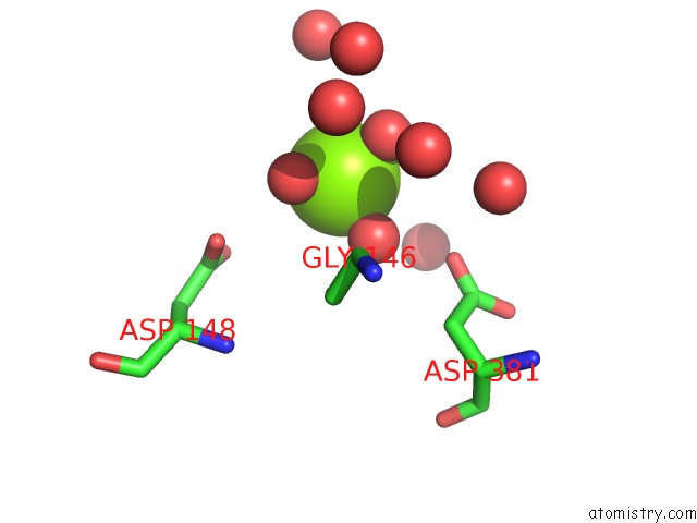
Mono view
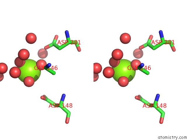
Stereo pair view
Mono view
Stereo pair view
A full contact list of Magnesium with other atoms in the Mg binding
site number 1 of Crystal Structure of Tyrosine Decarboxylase at 1.73 Angstroms Resolution within 5.0Å range:
|
Reference:
H.Zhu,
G.Xu,
K.Zhang,
X.Kong,
R.Han,
J.Zhou,
Y.Ni.
Crystal Structure of Tyrosine Decarboxylase and Identification of Key Residues Involved in Conformational Swing and Substrate Binding Sci Rep V. 6 27779 2016.
ISSN: ESSN 2045-2322
PubMed: 27292129
DOI: 10.1038/SREP27779
Page generated: Sun Sep 29 16:33:40 2024
ISSN: ESSN 2045-2322
PubMed: 27292129
DOI: 10.1038/SREP27779
Last articles
Mg in 5G0RMg in 5G5V
Mg in 5G3T
Mg in 5G5T
Mg in 5G5S
Mg in 5G4A
Mg in 5G57
Mg in 5G50
Mg in 5G41
Mg in 5G3Z