Magnesium »
PDB 5hr7-5i9e »
5hzh »
Magnesium in PDB 5hzh: Crystal Structure of Photoinhibitable RAC1 Containing C450A Mutant LOV2 Domain
Protein crystallography data
The structure of Crystal Structure of Photoinhibitable RAC1 Containing C450A Mutant LOV2 Domain, PDB code: 5hzh
was solved by
M.Tarnawski,
O.Dagliyan,
P.H.Chu,
D.Shirvanyants,
N.V.Dokholyan,
K.M.Hahn,
I.Schlichting,
with X-Ray Crystallography technique. A brief refinement statistics is given in the table below:
| Resolution Low / High (Å) | 37.14 / 2.60 |
| Space group | P 1 21 1 |
| Cell size a, b, c (Å), α, β, γ (°) | 36.050, 74.270, 58.400, 90.00, 94.62, 90.00 |
| R / Rfree (%) | 20.4 / 25.5 |
Other elements in 5hzh:
The structure of Crystal Structure of Photoinhibitable RAC1 Containing C450A Mutant LOV2 Domain also contains other interesting chemical elements:
| Calcium | (Ca) | 2 atoms |
Magnesium Binding Sites:
The binding sites of Magnesium atom in the Crystal Structure of Photoinhibitable RAC1 Containing C450A Mutant LOV2 Domain
(pdb code 5hzh). This binding sites where shown within
5.0 Angstroms radius around Magnesium atom.
In total only one binding site of Magnesium was determined in the Crystal Structure of Photoinhibitable RAC1 Containing C450A Mutant LOV2 Domain, PDB code: 5hzh:
In total only one binding site of Magnesium was determined in the Crystal Structure of Photoinhibitable RAC1 Containing C450A Mutant LOV2 Domain, PDB code: 5hzh:
Magnesium binding site 1 out of 1 in 5hzh
Go back to
Magnesium binding site 1 out
of 1 in the Crystal Structure of Photoinhibitable RAC1 Containing C450A Mutant LOV2 Domain

Mono view
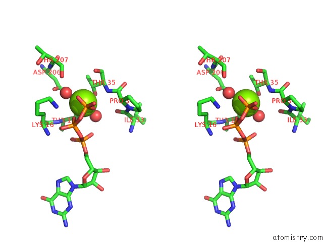
Stereo pair view
Mono view
Stereo pair view
A full contact list of Magnesium with other atoms in the Mg binding
site number 1 of Crystal Structure of Photoinhibitable RAC1 Containing C450A Mutant LOV2 Domain within 5.0Å range:
|
Reference:
O.Dagliyan,
M.Tarnawski,
P.H.Chu,
D.Shirvanyants,
I.Schlichting,
N.V.Dokholyan,
K.M.Hahn.
Engineering Extrinsic Disorder to Control Protein Activity in Living Cells. Science V. 354 1441 2016.
ISSN: ESSN 1095-9203
PubMed: 27980211
DOI: 10.1126/SCIENCE.AAH3404
Page generated: Sun Sep 29 16:37:27 2024
ISSN: ESSN 1095-9203
PubMed: 27980211
DOI: 10.1126/SCIENCE.AAH3404
Last articles
Mg in 3AUAMg in 3AU9
Mg in 3AU6
Mg in 3AU0
Mg in 3ATT
Mg in 3ATS
Mg in 3ARN
Mg in 3ATU
Mg in 3ASO
Mg in 3ATF