Magnesium »
PDB 5kty-5l5u »
5l0q »
Magnesium in PDB 5l0q: Crystal Structure of the Complex Between ADAM10 D+C Domain and A Conformation Specific Mab 8C7.
Enzymatic activity of Crystal Structure of the Complex Between ADAM10 D+C Domain and A Conformation Specific Mab 8C7.
All present enzymatic activity of Crystal Structure of the Complex Between ADAM10 D+C Domain and A Conformation Specific Mab 8C7.:
3.4.24.81;
3.4.24.81;
Protein crystallography data
The structure of Crystal Structure of the Complex Between ADAM10 D+C Domain and A Conformation Specific Mab 8C7., PDB code: 5l0q
was solved by
K.Xu,
N.Saha,
D.B.Nikolov,
with X-Ray Crystallography technique. A brief refinement statistics is given in the table below:
| Resolution Low / High (Å) | 125.26 / 2.76 |
| Space group | P 21 21 21 |
| Cell size a, b, c (Å), α, β, γ (°) | 53.326, 141.679, 268.080, 90.00, 90.00, 90.00 |
| R / Rfree (%) | 20.3 / 25.1 |
Magnesium Binding Sites:
The binding sites of Magnesium atom in the Crystal Structure of the Complex Between ADAM10 D+C Domain and A Conformation Specific Mab 8C7.
(pdb code 5l0q). This binding sites where shown within
5.0 Angstroms radius around Magnesium atom.
In total 2 binding sites of Magnesium where determined in the Crystal Structure of the Complex Between ADAM10 D+C Domain and A Conformation Specific Mab 8C7., PDB code: 5l0q:
Jump to Magnesium binding site number: 1; 2;
In total 2 binding sites of Magnesium where determined in the Crystal Structure of the Complex Between ADAM10 D+C Domain and A Conformation Specific Mab 8C7., PDB code: 5l0q:
Jump to Magnesium binding site number: 1; 2;
Magnesium binding site 1 out of 2 in 5l0q
Go back to
Magnesium binding site 1 out
of 2 in the Crystal Structure of the Complex Between ADAM10 D+C Domain and A Conformation Specific Mab 8C7.
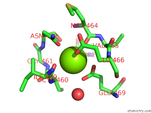
Mono view
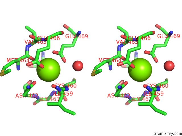
Stereo pair view
Mono view
Stereo pair view
A full contact list of Magnesium with other atoms in the Mg binding
site number 1 of Crystal Structure of the Complex Between ADAM10 D+C Domain and A Conformation Specific Mab 8C7. within 5.0Å range:
|
Magnesium binding site 2 out of 2 in 5l0q
Go back to
Magnesium binding site 2 out
of 2 in the Crystal Structure of the Complex Between ADAM10 D+C Domain and A Conformation Specific Mab 8C7.
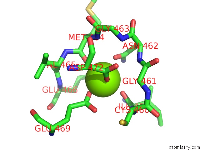
Mono view
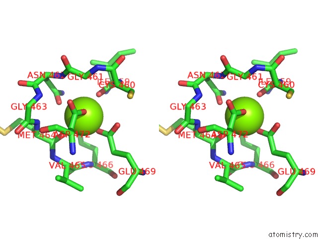
Stereo pair view
Mono view
Stereo pair view
A full contact list of Magnesium with other atoms in the Mg binding
site number 2 of Crystal Structure of the Complex Between ADAM10 D+C Domain and A Conformation Specific Mab 8C7. within 5.0Å range:
|
Reference:
L.Atapattu,
N.Saha,
C.Chheang,
M.F.Eissman,
K.Xu,
M.E.Vail,
L.Hii,
C.Llerena,
Z.Liu,
K.Horvay,
H.E.Abud,
U.Kusebauch,
R.L.Moritz,
B.S.Ding,
Z.Cao,
S.Rafii,
M.Ernst,
A.M.Scott,
D.B.Nikolov,
M.Lackmann,
P.W.Janes.
An Activated Form of ADAM10 Is Tumor Selective and Regulates Cancer Stem-Like Cells and Tumor Growth. J.Exp.Med. V. 213 1741 2016.
ISSN: ESSN 1540-9538
PubMed: 27503072
DOI: 10.1084/JEM.20151095
Page generated: Sun Sep 29 19:16:41 2024
ISSN: ESSN 1540-9538
PubMed: 27503072
DOI: 10.1084/JEM.20151095
Last articles
Mg in 5G0RMg in 5G5V
Mg in 5G3T
Mg in 5G5T
Mg in 5G5S
Mg in 5G4A
Mg in 5G57
Mg in 5G50
Mg in 5G41
Mg in 5G3Z