Magnesium »
PDB 5lu5-5m5i »
5m4l »
Magnesium in PDB 5m4l: Crystal Structure of Wild-Type Human Prolidase with Mg Ions and Leupro Ligand
Enzymatic activity of Crystal Structure of Wild-Type Human Prolidase with Mg Ions and Leupro Ligand
All present enzymatic activity of Crystal Structure of Wild-Type Human Prolidase with Mg Ions and Leupro Ligand:
3.4.13.9;
3.4.13.9;
Protein crystallography data
The structure of Crystal Structure of Wild-Type Human Prolidase with Mg Ions and Leupro Ligand, PDB code: 5m4l
was solved by
P.Wilk,
M.S.Weiss,
U.Mueller,
H.Dobbek,
with X-Ray Crystallography technique. A brief refinement statistics is given in the table below:
| Resolution Low / High (Å) | 46.75 / 1.49 |
| Space group | C 2 2 21 |
| Cell size a, b, c (Å), α, β, γ (°) | 103.605, 106.839, 216.990, 90.00, 90.00, 90.00 |
| R / Rfree (%) | 14.6 / 17.1 |
Other elements in 5m4l:
The structure of Crystal Structure of Wild-Type Human Prolidase with Mg Ions and Leupro Ligand also contains other interesting chemical elements:
| Sodium | (Na) | 1 atom |
Magnesium Binding Sites:
The binding sites of Magnesium atom in the Crystal Structure of Wild-Type Human Prolidase with Mg Ions and Leupro Ligand
(pdb code 5m4l). This binding sites where shown within
5.0 Angstroms radius around Magnesium atom.
In total 4 binding sites of Magnesium where determined in the Crystal Structure of Wild-Type Human Prolidase with Mg Ions and Leupro Ligand, PDB code: 5m4l:
Jump to Magnesium binding site number: 1; 2; 3; 4;
In total 4 binding sites of Magnesium where determined in the Crystal Structure of Wild-Type Human Prolidase with Mg Ions and Leupro Ligand, PDB code: 5m4l:
Jump to Magnesium binding site number: 1; 2; 3; 4;
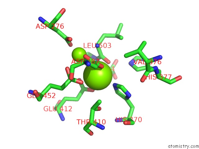
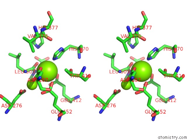
Magnesium binding site 1 out of 4 in 5m4l
Go back to
Magnesium binding site 1 out
of 4 in the Crystal Structure of Wild-Type Human Prolidase with Mg Ions and Leupro Ligand
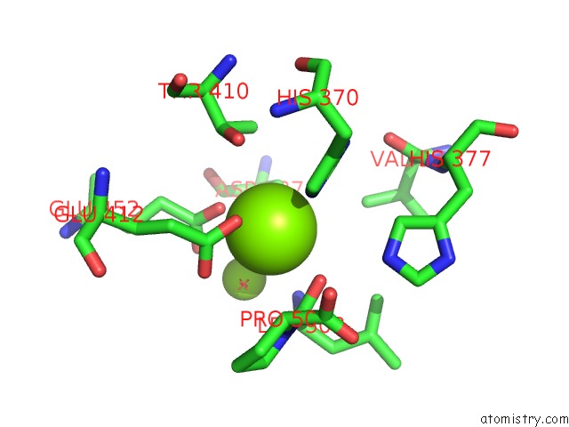
Mono view
Stereo pair view
Mono view
Stereo pair view
A full contact list of Magnesium with other atoms in the Mg binding
site number 1 of Crystal Structure of Wild-Type Human Prolidase with Mg Ions and Leupro Ligand within 5.0Å range:
|
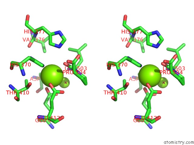
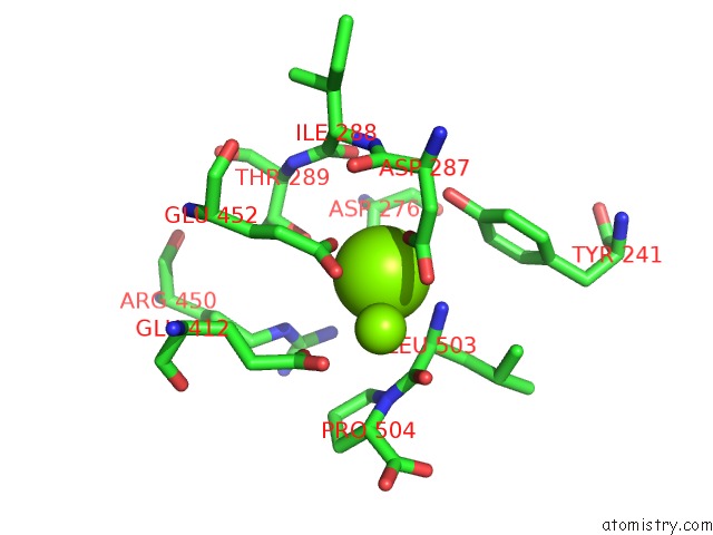
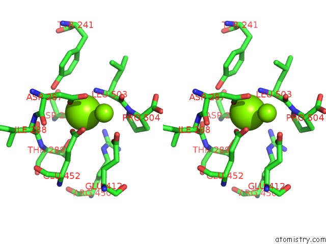
Magnesium binding site 2 out of 4 in 5m4l
Go back to
Magnesium binding site 2 out
of 4 in the Crystal Structure of Wild-Type Human Prolidase with Mg Ions and Leupro Ligand
Mono view
Stereo pair view
Mono view
Stereo pair view
A full contact list of Magnesium with other atoms in the Mg binding
site number 2 of Crystal Structure of Wild-Type Human Prolidase with Mg Ions and Leupro Ligand within 5.0Å range:
|
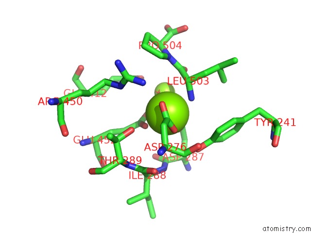
Magnesium binding site 3 out of 4 in 5m4l
Go back to
Magnesium binding site 3 out
of 4 in the Crystal Structure of Wild-Type Human Prolidase with Mg Ions and Leupro Ligand
Mono view
Stereo pair view
Mono view
Stereo pair view
A full contact list of Magnesium with other atoms in the Mg binding
site number 3 of Crystal Structure of Wild-Type Human Prolidase with Mg Ions and Leupro Ligand within 5.0Å range:
|
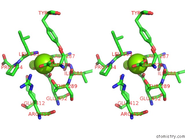
Magnesium binding site 4 out of 4 in 5m4l
Go back to
Magnesium binding site 4 out
of 4 in the Crystal Structure of Wild-Type Human Prolidase with Mg Ions and Leupro Ligand
Mono view
Stereo pair view
Mono view
Stereo pair view
A full contact list of Magnesium with other atoms in the Mg binding
site number 4 of Crystal Structure of Wild-Type Human Prolidase with Mg Ions and Leupro Ligand within 5.0Å range:
|
Reference:
P.Wilk,
M.Uehlein,
J.Kalms,
H.Dobbek,
U.Mueller,
M.S.Weiss.
Substrate Specificity and Reaction Mechanism of Human Prolidase. Febs J. V. 284 2870 2017.
ISSN: ISSN 1742-4658
PubMed: 28677335
DOI: 10.1111/FEBS.14158
Page generated: Tue Aug 12 14:49:05 2025
ISSN: ISSN 1742-4658
PubMed: 28677335
DOI: 10.1111/FEBS.14158
Last articles
Mn in 9LJUMn in 9LJW
Mn in 9LJS
Mn in 9LJR
Mn in 9LJT
Mn in 9LJV
Mg in 9UA2
Mg in 9R96
Mg in 9VM1
Mg in 9P01