Magnesium »
PDB 5o62-5odz »
5o69 »
Magnesium in PDB 5o69: The Structure of the Thermobifida Fusca Guanidine III Riboswitch with Agmatine.
Protein crystallography data
The structure of The Structure of the Thermobifida Fusca Guanidine III Riboswitch with Agmatine., PDB code: 5o69
was solved by
L.Huang,
J.Wang,
D.M.J.Lilley,
with X-Ray Crystallography technique. A brief refinement statistics is given in the table below:
| Resolution Low / High (Å) | 36.32 / 2.32 |
| Space group | P 31 2 1 |
| Cell size a, b, c (Å), α, β, γ (°) | 83.886, 83.886, 67.226, 90.00, 90.00, 120.00 |
| R / Rfree (%) | 24.3 / 27 |
Other elements in 5o69:
The structure of The Structure of the Thermobifida Fusca Guanidine III Riboswitch with Agmatine. also contains other interesting chemical elements:
| Bromine | (Br) | 4 atoms |
Magnesium Binding Sites:
The binding sites of Magnesium atom in the The Structure of the Thermobifida Fusca Guanidine III Riboswitch with Agmatine.
(pdb code 5o69). This binding sites where shown within
5.0 Angstroms radius around Magnesium atom.
In total only one binding site of Magnesium was determined in the The Structure of the Thermobifida Fusca Guanidine III Riboswitch with Agmatine., PDB code: 5o69:
In total only one binding site of Magnesium was determined in the The Structure of the Thermobifida Fusca Guanidine III Riboswitch with Agmatine., PDB code: 5o69:
Magnesium binding site 1 out of 1 in 5o69
Go back to
Magnesium binding site 1 out
of 1 in the The Structure of the Thermobifida Fusca Guanidine III Riboswitch with Agmatine.
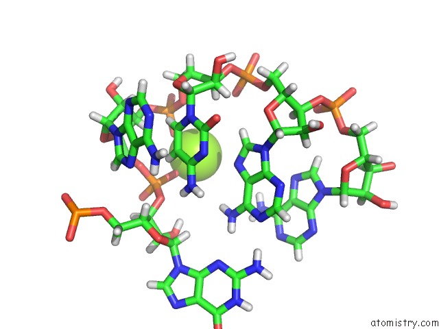
Mono view
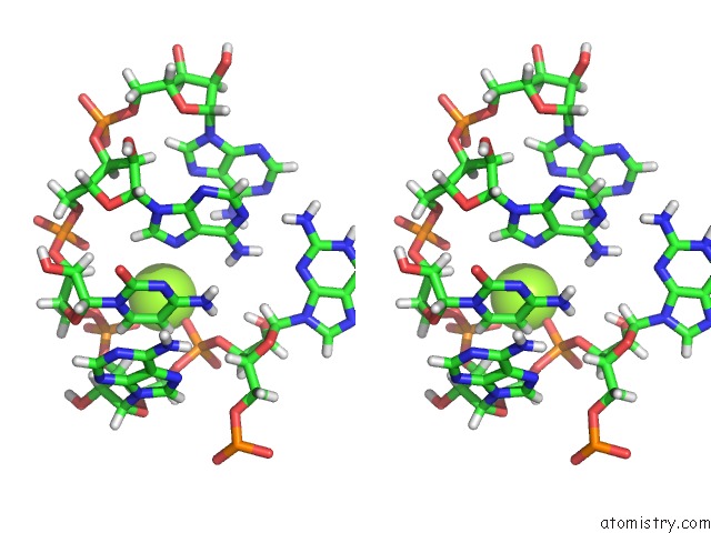
Stereo pair view
Mono view
Stereo pair view
A full contact list of Magnesium with other atoms in the Mg binding
site number 1 of The Structure of the Thermobifida Fusca Guanidine III Riboswitch with Agmatine. within 5.0Å range:
|
Reference:
L.Huang,
J.Wang,
T.J.Wilson,
D.M.J.Lilley.
Structure of the Guanidine III Riboswitch. Cell Chem Biol V. 24 1407 2017.
ISSN: ESSN 2451-9456
PubMed: 28988949
DOI: 10.1016/J.CHEMBIOL.2017.08.021
Page generated: Mon Sep 30 00:11:43 2024
ISSN: ESSN 2451-9456
PubMed: 28988949
DOI: 10.1016/J.CHEMBIOL.2017.08.021
Last articles
Mg in 4DUYMg in 4DR7
Mg in 4DR6
Mg in 4DR5
Mg in 4DUX
Mg in 4DUW
Mg in 4DUV
Mg in 4DUO
Mg in 4DUG
Mg in 4DTY