Magnesium »
PDB 5o62-5odz »
5odo »
Magnesium in PDB 5odo: Crystal Structure of the Oleate Hydratase of Rhodococcus Erythropolis
Protein crystallography data
The structure of Crystal Structure of the Oleate Hydratase of Rhodococcus Erythropolis, PDB code: 5odo
was solved by
R.Driller,
J.Lorenzen,
A.Waldow,
F.Qoura,
T.Brueck,
B.Loll,
with X-Ray Crystallography technique. A brief refinement statistics is given in the table below:
| Resolution Low / High (Å) | 49.47 / 2.64 |
| Space group | P 65 2 2 |
| Cell size a, b, c (Å), α, β, γ (°) | 261.747, 261.747, 127.304, 90.00, 90.00, 120.00 |
| R / Rfree (%) | 18.9 / 22 |
Magnesium Binding Sites:
The binding sites of Magnesium atom in the Crystal Structure of the Oleate Hydratase of Rhodococcus Erythropolis
(pdb code 5odo). This binding sites where shown within
5.0 Angstroms radius around Magnesium atom.
In total 2 binding sites of Magnesium where determined in the Crystal Structure of the Oleate Hydratase of Rhodococcus Erythropolis, PDB code: 5odo:
Jump to Magnesium binding site number: 1; 2;
In total 2 binding sites of Magnesium where determined in the Crystal Structure of the Oleate Hydratase of Rhodococcus Erythropolis, PDB code: 5odo:
Jump to Magnesium binding site number: 1; 2;
Magnesium binding site 1 out of 2 in 5odo
Go back to
Magnesium binding site 1 out
of 2 in the Crystal Structure of the Oleate Hydratase of Rhodococcus Erythropolis
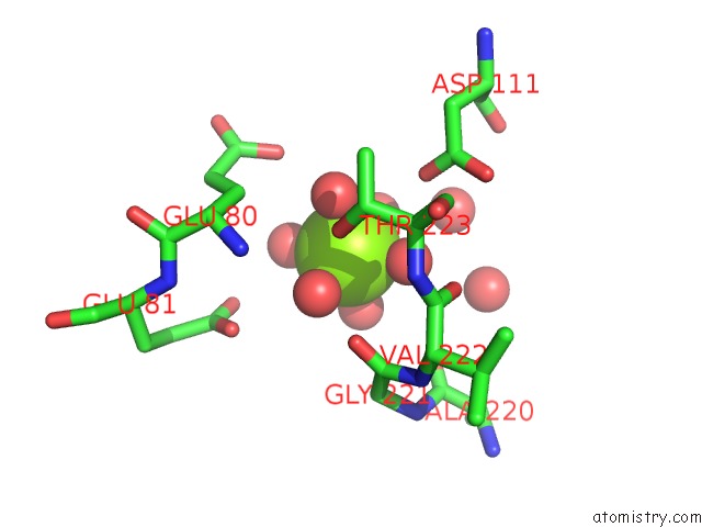
Mono view
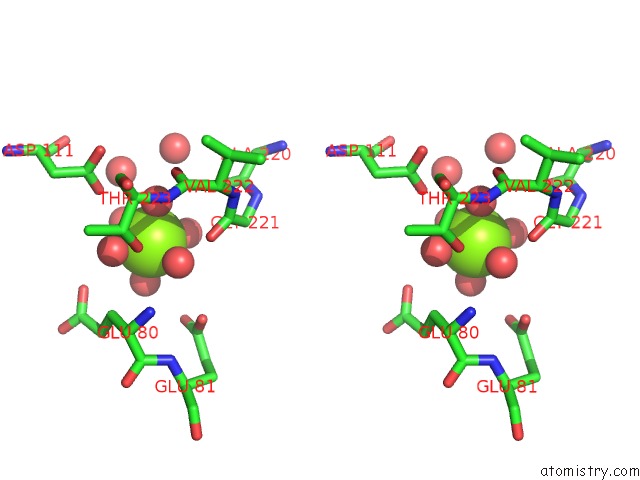
Stereo pair view
Mono view
Stereo pair view
A full contact list of Magnesium with other atoms in the Mg binding
site number 1 of Crystal Structure of the Oleate Hydratase of Rhodococcus Erythropolis within 5.0Å range:
|
Magnesium binding site 2 out of 2 in 5odo
Go back to
Magnesium binding site 2 out
of 2 in the Crystal Structure of the Oleate Hydratase of Rhodococcus Erythropolis
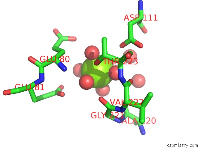
Mono view
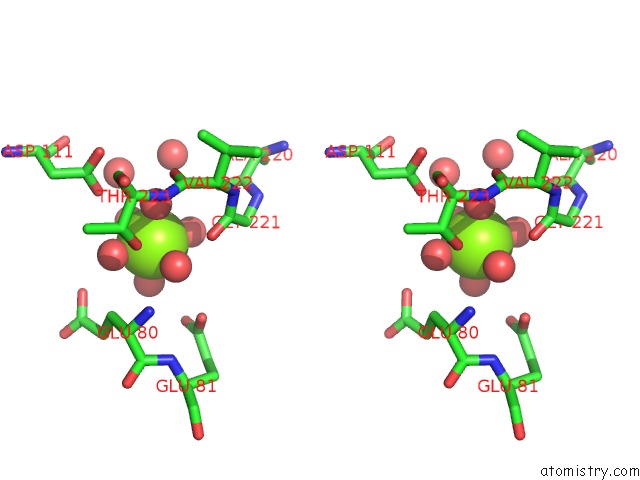
Stereo pair view
Mono view
Stereo pair view
A full contact list of Magnesium with other atoms in the Mg binding
site number 2 of Crystal Structure of the Oleate Hydratase of Rhodococcus Erythropolis within 5.0Å range:
|
Reference:
J.Lorenzen,
R.Driller,
A.Waldow,
F.Quora,
B.Loll,
T.Brueck.
Rhodococcus Erythropolis Oleate Hydratase: A New Member in the Oleate Hydratase Family Tree - Biochemical and Structural Studies. Chemcatchem 2017.
ISSN: ESSN 1867-3899
DOI: 10.1002/CCTC.201701350R1
Page generated: Mon Sep 30 00:18:57 2024
ISSN: ESSN 1867-3899
DOI: 10.1002/CCTC.201701350R1
Last articles
Mg in 4DR7Mg in 4DR6
Mg in 4DR5
Mg in 4DUX
Mg in 4DUW
Mg in 4DUV
Mg in 4DUO
Mg in 4DUG
Mg in 4DTY
Mg in 4DTW