Magnesium »
PDB 5t3k-5tfc »
5td3 »
Magnesium in PDB 5td3: Crystal Structure of Catechol 1,2-Dioxygenase From Burkholderia Vietnamiensis
Enzymatic activity of Crystal Structure of Catechol 1,2-Dioxygenase From Burkholderia Vietnamiensis
All present enzymatic activity of Crystal Structure of Catechol 1,2-Dioxygenase From Burkholderia Vietnamiensis:
1.13.11.1;
1.13.11.1;
Protein crystallography data
The structure of Crystal Structure of Catechol 1,2-Dioxygenase From Burkholderia Vietnamiensis, PDB code: 5td3
was solved by
Seattle Structural Genomics Center For Infectious Disease (Ssgcid),
with X-Ray Crystallography technique. A brief refinement statistics is given in the table below:
| Resolution Low / High (Å) | 40.51 / 1.75 |
| Space group | C 1 2 1 |
| Cell size a, b, c (Å), α, β, γ (°) | 169.410, 55.860, 85.690, 90.00, 116.30, 90.00 |
| R / Rfree (%) | 17.7 / 21 |
Other elements in 5td3:
The structure of Crystal Structure of Catechol 1,2-Dioxygenase From Burkholderia Vietnamiensis also contains other interesting chemical elements:
| Zinc | (Zn) | 2 atoms |
| Chlorine | (Cl) | 2 atoms |
Magnesium Binding Sites:
The binding sites of Magnesium atom in the Crystal Structure of Catechol 1,2-Dioxygenase From Burkholderia Vietnamiensis
(pdb code 5td3). This binding sites where shown within
5.0 Angstroms radius around Magnesium atom.
In total only one binding site of Magnesium was determined in the Crystal Structure of Catechol 1,2-Dioxygenase From Burkholderia Vietnamiensis, PDB code: 5td3:
In total only one binding site of Magnesium was determined in the Crystal Structure of Catechol 1,2-Dioxygenase From Burkholderia Vietnamiensis, PDB code: 5td3:
Magnesium binding site 1 out of 1 in 5td3
Go back to
Magnesium binding site 1 out
of 1 in the Crystal Structure of Catechol 1,2-Dioxygenase From Burkholderia Vietnamiensis
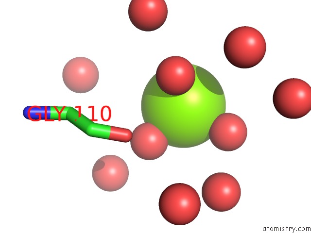
Mono view
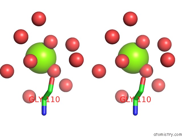
Stereo pair view
Mono view
Stereo pair view
A full contact list of Magnesium with other atoms in the Mg binding
site number 1 of Crystal Structure of Catechol 1,2-Dioxygenase From Burkholderia Vietnamiensis within 5.0Å range:
|
Reference:
D.G.Conrady,
D.M.Dranow,
D.Lorimer,
T.E.Edwards.
Crystal Structure of Catechol 1,2-Dioxygenase From Burkholderia Vietnamiensis To Be Published.
Page generated: Mon Sep 30 04:48:22 2024
Last articles
K in 5FKFK in 5FK6
K in 5FKE
K in 5FKD
K in 5FK5
K in 5FJC
K in 5FK3
K in 5FHW
K in 5FCW
K in 5FG0