Magnesium »
PDB 5tfd-5tus »
5thk »
Magnesium in PDB 5thk: Crystal Structure of A Putative Dehydrogenase From Burkholderia Cenocepacia with Bound Nadp
Protein crystallography data
The structure of Crystal Structure of A Putative Dehydrogenase From Burkholderia Cenocepacia with Bound Nadp, PDB code: 5thk
was solved by
Seattle Structural Genomics Center For Infectious Disease (Ssgcid),
with X-Ray Crystallography technique. A brief refinement statistics is given in the table below:
| Resolution Low / High (Å) | 45.19 / 1.40 |
| Space group | P 1 21 1 |
| Cell size a, b, c (Å), α, β, γ (°) | 75.540, 102.440, 122.250, 90.00, 97.28, 90.00 |
| R / Rfree (%) | 14 / 16.4 |
Magnesium Binding Sites:
The binding sites of Magnesium atom in the Crystal Structure of A Putative Dehydrogenase From Burkholderia Cenocepacia with Bound Nadp
(pdb code 5thk). This binding sites where shown within
5.0 Angstroms radius around Magnesium atom.
In total 2 binding sites of Magnesium where determined in the Crystal Structure of A Putative Dehydrogenase From Burkholderia Cenocepacia with Bound Nadp, PDB code: 5thk:
Jump to Magnesium binding site number: 1; 2;
In total 2 binding sites of Magnesium where determined in the Crystal Structure of A Putative Dehydrogenase From Burkholderia Cenocepacia with Bound Nadp, PDB code: 5thk:
Jump to Magnesium binding site number: 1; 2;
Magnesium binding site 1 out of 2 in 5thk
Go back to
Magnesium binding site 1 out
of 2 in the Crystal Structure of A Putative Dehydrogenase From Burkholderia Cenocepacia with Bound Nadp
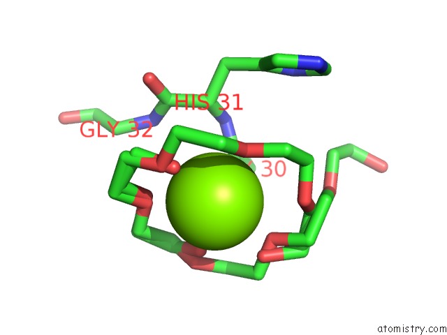
Mono view
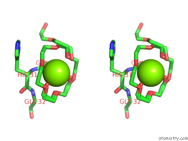
Stereo pair view
Mono view
Stereo pair view
A full contact list of Magnesium with other atoms in the Mg binding
site number 1 of Crystal Structure of A Putative Dehydrogenase From Burkholderia Cenocepacia with Bound Nadp within 5.0Å range:
|
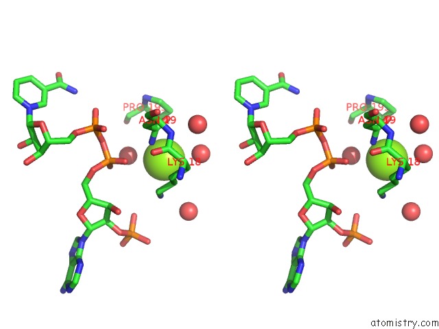
Magnesium binding site 2 out of 2 in 5thk
Go back to
Magnesium binding site 2 out
of 2 in the Crystal Structure of A Putative Dehydrogenase From Burkholderia Cenocepacia with Bound Nadp
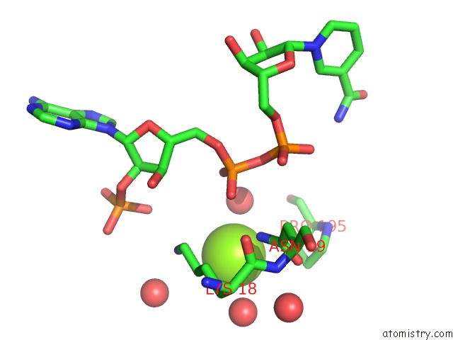
Mono view
Stereo pair view
Mono view
Stereo pair view
A full contact list of Magnesium with other atoms in the Mg binding
site number 2 of Crystal Structure of A Putative Dehydrogenase From Burkholderia Cenocepacia with Bound Nadp within 5.0Å range:
|
Reference:
D.M.Dranow,
S.J.Mayclin,
D.D.Lorimer,
T.E.Edwards.
Crystal Structure of A Putative Dehydrogenase From Burkholderia Cenocepacia with Bound Nadp To Be Published.
Page generated: Mon Sep 30 04:51:50 2024
Last articles
Mg in 5HR6Mg in 5HQL
Mg in 5HQW
Mg in 5HQM
Mg in 5HQC
Mg in 5HQB
Mg in 5HQ8
Mg in 5HQA
Mg in 5HQ4
Mg in 5HPY