Magnesium »
PDB 5u7d-5uhd »
5ucr »
Magnesium in PDB 5ucr: Crystal Structure of A Pantoate-Beta-Alanine Ligase From Neisseria Gonorrhoeae with Bound Amppnp and Alanine
Enzymatic activity of Crystal Structure of A Pantoate-Beta-Alanine Ligase From Neisseria Gonorrhoeae with Bound Amppnp and Alanine
All present enzymatic activity of Crystal Structure of A Pantoate-Beta-Alanine Ligase From Neisseria Gonorrhoeae with Bound Amppnp and Alanine:
6.3.2.1;
6.3.2.1;
Protein crystallography data
The structure of Crystal Structure of A Pantoate-Beta-Alanine Ligase From Neisseria Gonorrhoeae with Bound Amppnp and Alanine, PDB code: 5ucr
was solved by
Seattle Structural Genomics Center For Infectious Disease,
Seattlestructural Genomics Center For Infectious Disease (Ssgcid),
with X-Ray Crystallography technique. A brief refinement statistics is given in the table below:
| Resolution Low / High (Å) | 40.49 / 2.25 |
| Space group | P 41 21 2 |
| Cell size a, b, c (Å), α, β, γ (°) | 137.650, 137.650, 72.950, 90.00, 90.00, 90.00 |
| R / Rfree (%) | 17.4 / 21.7 |
Magnesium Binding Sites:
The binding sites of Magnesium atom in the Crystal Structure of A Pantoate-Beta-Alanine Ligase From Neisseria Gonorrhoeae with Bound Amppnp and Alanine
(pdb code 5ucr). This binding sites where shown within
5.0 Angstroms radius around Magnesium atom.
In total only one binding site of Magnesium was determined in the Crystal Structure of A Pantoate-Beta-Alanine Ligase From Neisseria Gonorrhoeae with Bound Amppnp and Alanine, PDB code: 5ucr:
In total only one binding site of Magnesium was determined in the Crystal Structure of A Pantoate-Beta-Alanine Ligase From Neisseria Gonorrhoeae with Bound Amppnp and Alanine, PDB code: 5ucr:
Magnesium binding site 1 out of 1 in 5ucr
Go back to
Magnesium binding site 1 out
of 1 in the Crystal Structure of A Pantoate-Beta-Alanine Ligase From Neisseria Gonorrhoeae with Bound Amppnp and Alanine
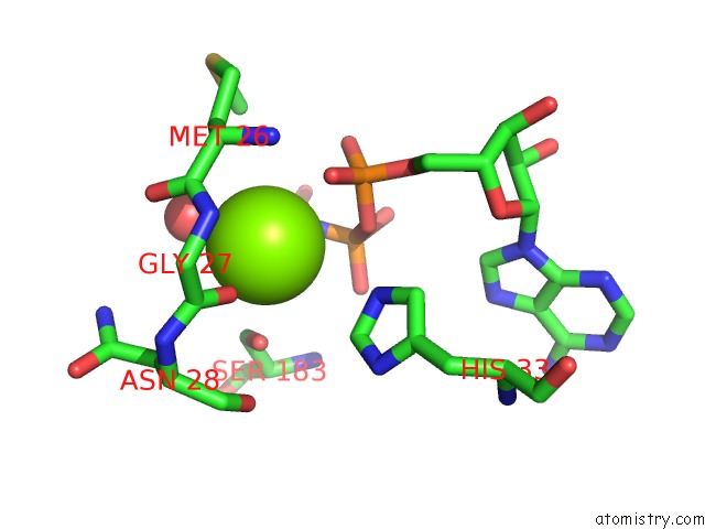
Mono view
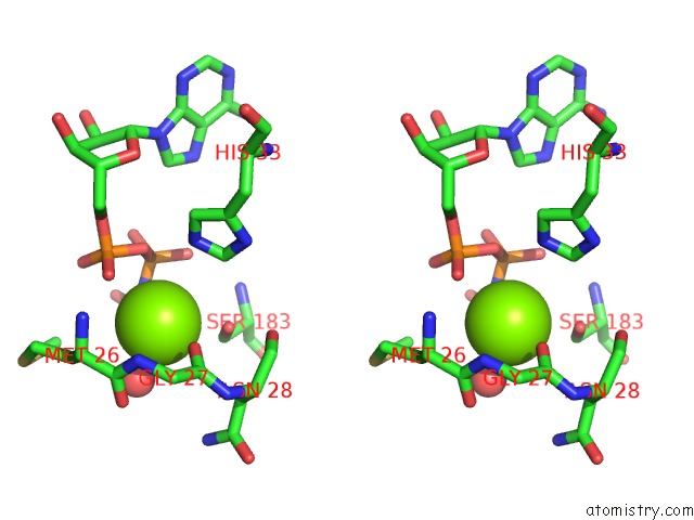
Stereo pair view
Mono view
Stereo pair view
A full contact list of Magnesium with other atoms in the Mg binding
site number 1 of Crystal Structure of A Pantoate-Beta-Alanine Ligase From Neisseria Gonorrhoeae with Bound Amppnp and Alanine within 5.0Å range:
|
Reference:
D.M.Dranow,
S.J.Mayclin,
D.D.Lorimer,
T.E.Edwards.
Crystal Structure of A Pantoate-Beta-Alanine Ligase From Neisseria Gonorrhoeae with Bound Amppnp and Alanine To Be Published.
Page generated: Tue Aug 12 20:48:00 2025
Last articles
Mg in 6CKXMg in 6CK4
Mg in 6CKB
Mg in 6CK5
Mg in 6CJJ
Mg in 6CJL
Mg in 6CJ4
Mg in 6CJ3
Mg in 6CJ0
Mg in 6CIP