Magnesium »
PDB 5vwa-5w51 »
5vz0 »
Magnesium in PDB 5vz0: Crystal Structure of Lactococcus Lactis Pyruvate Carboxylase G746A Mutant in Complex with Cyclic-Di-Amp
Enzymatic activity of Crystal Structure of Lactococcus Lactis Pyruvate Carboxylase G746A Mutant in Complex with Cyclic-Di-Amp
All present enzymatic activity of Crystal Structure of Lactococcus Lactis Pyruvate Carboxylase G746A Mutant in Complex with Cyclic-Di-Amp:
6.4.1.1;
6.4.1.1;
Protein crystallography data
The structure of Crystal Structure of Lactococcus Lactis Pyruvate Carboxylase G746A Mutant in Complex with Cyclic-Di-Amp, PDB code: 5vz0
was solved by
P.H.Choi,
L.Tong,
with X-Ray Crystallography technique. A brief refinement statistics is given in the table below:
| Resolution Low / High (Å) | 48.55 / 2.00 |
| Space group | P 1 |
| Cell size a, b, c (Å), α, β, γ (°) | 97.155, 130.390, 134.271, 65.99, 88.67, 70.14 |
| R / Rfree (%) | 19.1 / 21.8 |
Other elements in 5vz0:
The structure of Crystal Structure of Lactococcus Lactis Pyruvate Carboxylase G746A Mutant in Complex with Cyclic-Di-Amp also contains other interesting chemical elements:
| Manganese | (Mn) | 4 atoms |
Magnesium Binding Sites:
The binding sites of Magnesium atom in the Crystal Structure of Lactococcus Lactis Pyruvate Carboxylase G746A Mutant in Complex with Cyclic-Di-Amp
(pdb code 5vz0). This binding sites where shown within
5.0 Angstroms radius around Magnesium atom.
In total 3 binding sites of Magnesium where determined in the Crystal Structure of Lactococcus Lactis Pyruvate Carboxylase G746A Mutant in Complex with Cyclic-Di-Amp, PDB code: 5vz0:
Jump to Magnesium binding site number: 1; 2; 3;
In total 3 binding sites of Magnesium where determined in the Crystal Structure of Lactococcus Lactis Pyruvate Carboxylase G746A Mutant in Complex with Cyclic-Di-Amp, PDB code: 5vz0:
Jump to Magnesium binding site number: 1; 2; 3;
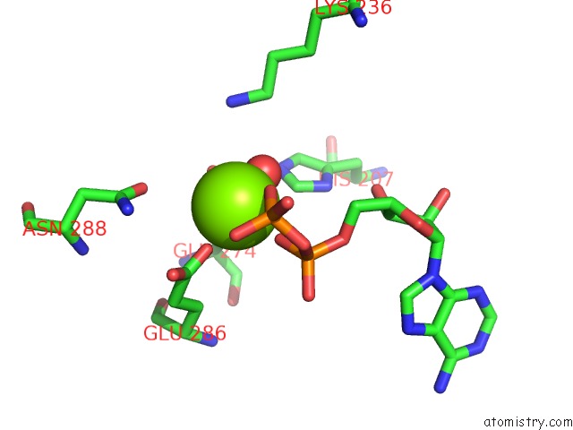
Magnesium binding site 1 out of 3 in 5vz0
Go back to
Magnesium binding site 1 out
of 3 in the Crystal Structure of Lactococcus Lactis Pyruvate Carboxylase G746A Mutant in Complex with Cyclic-Di-Amp
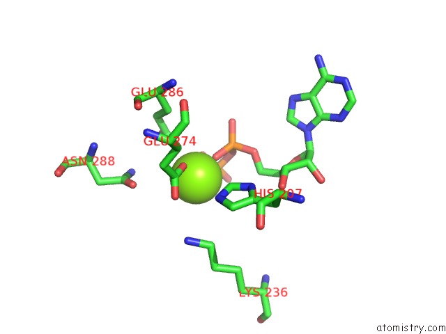
Mono view
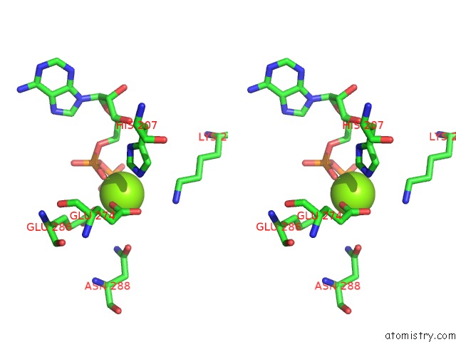
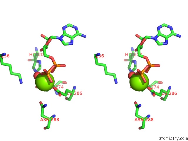
Stereo pair view
Mono view
Stereo pair view
A full contact list of Magnesium with other atoms in the Mg binding
site number 1 of Crystal Structure of Lactococcus Lactis Pyruvate Carboxylase G746A Mutant in Complex with Cyclic-Di-Amp within 5.0Å range:
|
Magnesium binding site 2 out of 3 in 5vz0
Go back to
Magnesium binding site 2 out
of 3 in the Crystal Structure of Lactococcus Lactis Pyruvate Carboxylase G746A Mutant in Complex with Cyclic-Di-Amp
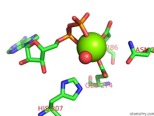
Mono view
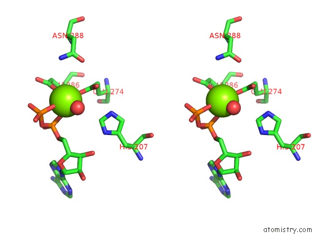
Stereo pair view
Mono view
Stereo pair view
A full contact list of Magnesium with other atoms in the Mg binding
site number 2 of Crystal Structure of Lactococcus Lactis Pyruvate Carboxylase G746A Mutant in Complex with Cyclic-Di-Amp within 5.0Å range:
|
Magnesium binding site 3 out of 3 in 5vz0
Go back to
Magnesium binding site 3 out
of 3 in the Crystal Structure of Lactococcus Lactis Pyruvate Carboxylase G746A Mutant in Complex with Cyclic-Di-Amp
Mono view
Stereo pair view
Mono view
Stereo pair view
A full contact list of Magnesium with other atoms in the Mg binding
site number 3 of Crystal Structure of Lactococcus Lactis Pyruvate Carboxylase G746A Mutant in Complex with Cyclic-Di-Amp within 5.0Å range:
|
Reference:
P.H.Choi,
T.M.N.Vu,
H.T.Pham,
J.J.Woodward,
M.S.Turner,
L.Tong.
Structural and Functional Studies of Pyruvate Carboxylase Regulation By Cyclic Di-Amp in Lactic Acid Bacteria. Proc. Natl. Acad. Sci. V. 114 E7226 2017U.S.A..
ISSN: ESSN 1091-6490
PubMed: 28808024
DOI: 10.1073/PNAS.1704756114
Page generated: Mon Sep 30 06:22:52 2024
ISSN: ESSN 1091-6490
PubMed: 28808024
DOI: 10.1073/PNAS.1704756114
Last articles
K in 6AQPK in 6B2W
K in 6B10
K in 6AI6
K in 6AU4
K in 6ASO
K in 6AFZ
K in 6AFY
K in 6AFX
K in 6AFW