Magnesium »
PDB 5wu4-5x8a »
5x0i »
Magnesium in PDB 5x0i: Crystal Structure of PKM2 R399E Mutant Complexed with Fbp and Serine
Enzymatic activity of Crystal Structure of PKM2 R399E Mutant Complexed with Fbp and Serine
All present enzymatic activity of Crystal Structure of PKM2 R399E Mutant Complexed with Fbp and Serine:
2.7.1.40;
2.7.1.40;
Protein crystallography data
The structure of Crystal Structure of PKM2 R399E Mutant Complexed with Fbp and Serine, PDB code: 5x0i
was solved by
W.C.Wang,
T.J.Chen,
with X-Ray Crystallography technique. A brief refinement statistics is given in the table below:
| Resolution Low / High (Å) | 30.00 / 2.64 |
| Space group | P 21 21 21 |
| Cell size a, b, c (Å), α, β, γ (°) | 116.601, 137.875, 149.709, 90.00, 90.00, 90.00 |
| R / Rfree (%) | 18 / 21.8 |
Other elements in 5x0i:
The structure of Crystal Structure of PKM2 R399E Mutant Complexed with Fbp and Serine also contains other interesting chemical elements:
| Potassium | (K) | 4 atoms |
Magnesium Binding Sites:
The binding sites of Magnesium atom in the Crystal Structure of PKM2 R399E Mutant Complexed with Fbp and Serine
(pdb code 5x0i). This binding sites where shown within
5.0 Angstroms radius around Magnesium atom.
In total 4 binding sites of Magnesium where determined in the Crystal Structure of PKM2 R399E Mutant Complexed with Fbp and Serine, PDB code: 5x0i:
Jump to Magnesium binding site number: 1; 2; 3; 4;
In total 4 binding sites of Magnesium where determined in the Crystal Structure of PKM2 R399E Mutant Complexed with Fbp and Serine, PDB code: 5x0i:
Jump to Magnesium binding site number: 1; 2; 3; 4;
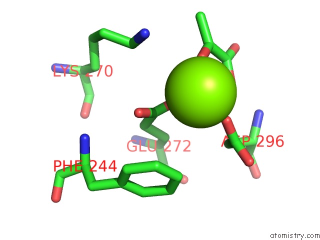
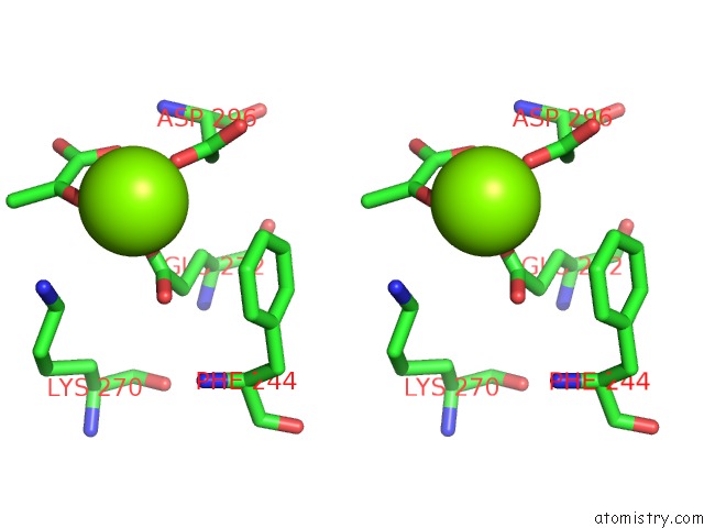
Magnesium binding site 1 out of 4 in 5x0i
Go back to
Magnesium binding site 1 out
of 4 in the Crystal Structure of PKM2 R399E Mutant Complexed with Fbp and Serine
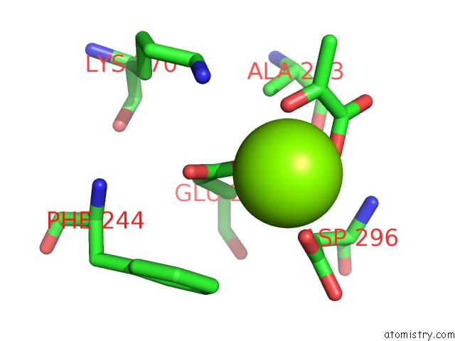
Mono view
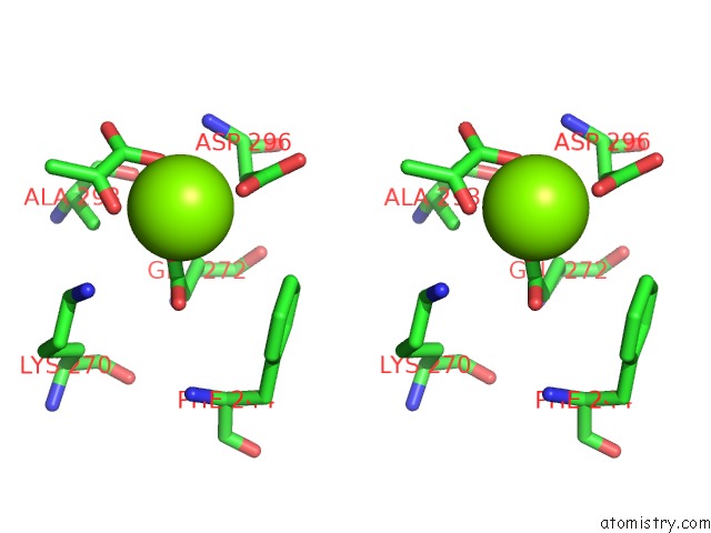
Stereo pair view
Mono view
Stereo pair view
A full contact list of Magnesium with other atoms in the Mg binding
site number 1 of Crystal Structure of PKM2 R399E Mutant Complexed with Fbp and Serine within 5.0Å range:
|
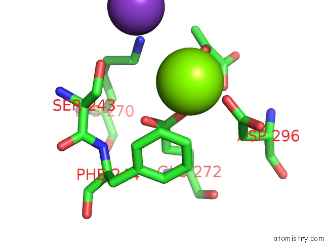
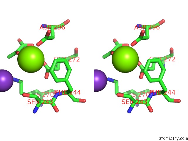
Magnesium binding site 2 out of 4 in 5x0i
Go back to
Magnesium binding site 2 out
of 4 in the Crystal Structure of PKM2 R399E Mutant Complexed with Fbp and Serine
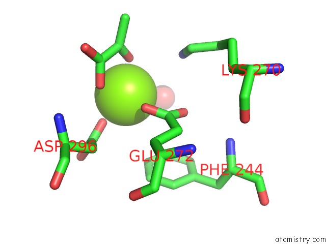
Mono view
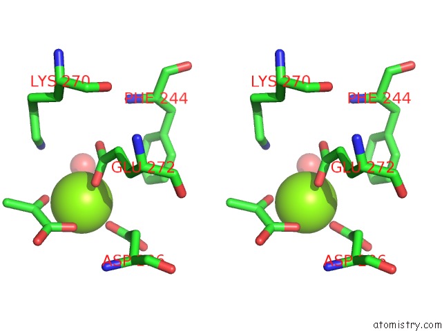
Stereo pair view
Mono view
Stereo pair view
A full contact list of Magnesium with other atoms in the Mg binding
site number 2 of Crystal Structure of PKM2 R399E Mutant Complexed with Fbp and Serine within 5.0Å range:
|
Magnesium binding site 3 out of 4 in 5x0i
Go back to
Magnesium binding site 3 out
of 4 in the Crystal Structure of PKM2 R399E Mutant Complexed with Fbp and Serine
Mono view
Stereo pair view
Mono view
Stereo pair view
A full contact list of Magnesium with other atoms in the Mg binding
site number 3 of Crystal Structure of PKM2 R399E Mutant Complexed with Fbp and Serine within 5.0Å range:
|
Magnesium binding site 4 out of 4 in 5x0i
Go back to
Magnesium binding site 4 out
of 4 in the Crystal Structure of PKM2 R399E Mutant Complexed with Fbp and Serine
Mono view
Stereo pair view
Mono view
Stereo pair view
A full contact list of Magnesium with other atoms in the Mg binding
site number 4 of Crystal Structure of PKM2 R399E Mutant Complexed with Fbp and Serine within 5.0Å range:
|
Reference:
T.J.Chen,
H.J.Wang,
J.S.Liu,
H.H.Cheng,
S.C.Hsu,
M.C.Wu,
C.H.Lu,
Y.F.Wu,
J.W.Wu,
Y.Y.Liu,
H.J.Kung,
W.C.Wang.
Mutations in the PKM2 Exon-10 Region Are Associated with Reduced Allostery and Increased Nuclear Translocation. Commun Biol V. 2 105 2019.
ISSN: ESSN 2399-3642
PubMed: 30911680
DOI: 10.1038/S42003-019-0343-4
Page generated: Tue Aug 12 23:16:06 2025
ISSN: ESSN 2399-3642
PubMed: 30911680
DOI: 10.1038/S42003-019-0343-4
Last articles
Mg in 6XU1Mg in 6Y0Z
Mg in 6Y0T
Mg in 6Y0Y
Mg in 6Y09
Mg in 6XYD
Mg in 6XYB
Mg in 6XY5
Mg in 6XXC
Mg in 6XXQ