Magnesium »
PDB 5y5p-5ylv »
5y5q »
Magnesium in PDB 5y5q: Crystal Structure of the Wssv Dutpase D88N/R158E Mutant in Complex with Dutp
Protein crystallography data
The structure of Crystal Structure of the Wssv Dutpase D88N/R158E Mutant in Complex with Dutp, PDB code: 5y5q
was solved by
Q.Ma,
K.Zang,
with X-Ray Crystallography technique. A brief refinement statistics is given in the table below:
| Resolution Low / High (Å) | 22.84 / 1.56 |
| Space group | P 21 21 21 |
| Cell size a, b, c (Å), α, β, γ (°) | 59.574, 75.527, 113.321, 90.00, 90.00, 90.00 |
| R / Rfree (%) | 17.3 / 19.2 |
Magnesium Binding Sites:
The binding sites of Magnesium atom in the Crystal Structure of the Wssv Dutpase D88N/R158E Mutant in Complex with Dutp
(pdb code 5y5q). This binding sites where shown within
5.0 Angstroms radius around Magnesium atom.
In total 3 binding sites of Magnesium where determined in the Crystal Structure of the Wssv Dutpase D88N/R158E Mutant in Complex with Dutp, PDB code: 5y5q:
Jump to Magnesium binding site number: 1; 2; 3;
In total 3 binding sites of Magnesium where determined in the Crystal Structure of the Wssv Dutpase D88N/R158E Mutant in Complex with Dutp, PDB code: 5y5q:
Jump to Magnesium binding site number: 1; 2; 3;
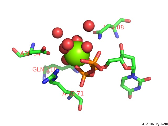
Magnesium binding site 1 out of 3 in 5y5q
Go back to
Magnesium binding site 1 out
of 3 in the Crystal Structure of the Wssv Dutpase D88N/R158E Mutant in Complex with Dutp
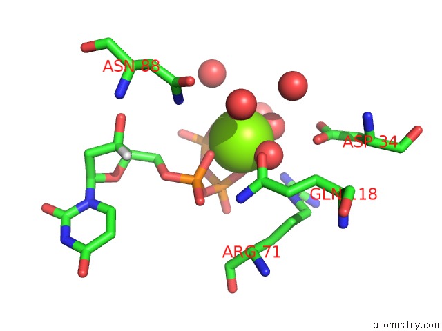
Mono view
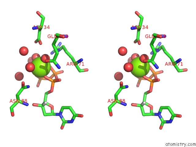
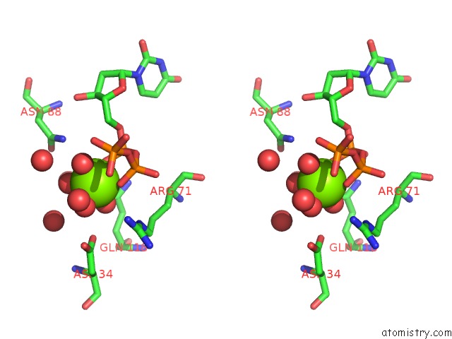
Stereo pair view
Mono view
Stereo pair view
A full contact list of Magnesium with other atoms in the Mg binding
site number 1 of Crystal Structure of the Wssv Dutpase D88N/R158E Mutant in Complex with Dutp within 5.0Å range:
|
Magnesium binding site 2 out of 3 in 5y5q
Go back to
Magnesium binding site 2 out
of 3 in the Crystal Structure of the Wssv Dutpase D88N/R158E Mutant in Complex with Dutp
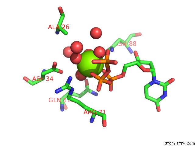
Mono view
Stereo pair view
Mono view
Stereo pair view
A full contact list of Magnesium with other atoms in the Mg binding
site number 2 of Crystal Structure of the Wssv Dutpase D88N/R158E Mutant in Complex with Dutp within 5.0Å range:
|
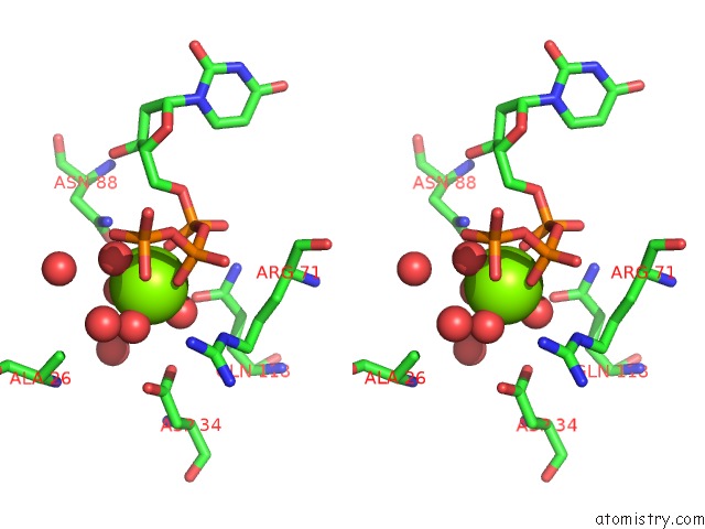
Magnesium binding site 3 out of 3 in 5y5q
Go back to
Magnesium binding site 3 out
of 3 in the Crystal Structure of the Wssv Dutpase D88N/R158E Mutant in Complex with Dutp
Mono view
Stereo pair view
Mono view
Stereo pair view
A full contact list of Magnesium with other atoms in the Mg binding
site number 3 of Crystal Structure of the Wssv Dutpase D88N/R158E Mutant in Complex with Dutp within 5.0Å range:
|
Reference:
K.Zang,
F.Li,
Q.Ma.
The Dutpase of White Spot Syndrome Virus Assembles Its Active Sites in A Noncanonical Manner. J. Biol. Chem. V. 293 1088 2018.
ISSN: ESSN 1083-351X
PubMed: 29187596
DOI: 10.1074/JBC.M117.815266
Page generated: Wed Aug 13 00:45:08 2025
ISSN: ESSN 1083-351X
PubMed: 29187596
DOI: 10.1074/JBC.M117.815266
Last articles
Na in 4RA5Na in 4R9I
Na in 4R8I
Na in 4R6C
Na in 4R6K
Na in 4R66
Na in 4R63
Na in 4R65
Na in 4R64
Na in 4R60