Magnesium »
PDB 5zkj-6a1l »
5zyd »
Magnesium in PDB 5zyd: Crystal Structure of Glucose Isomerase Soaked with Glucose
Enzymatic activity of Crystal Structure of Glucose Isomerase Soaked with Glucose
All present enzymatic activity of Crystal Structure of Glucose Isomerase Soaked with Glucose:
5.3.1.5;
5.3.1.5;
Protein crystallography data
The structure of Crystal Structure of Glucose Isomerase Soaked with Glucose, PDB code: 5zyd
was solved by
K.H.Nam,
with X-Ray Crystallography technique. A brief refinement statistics is given in the table below:
| Resolution Low / High (Å) | 70.70 / 1.40 |
| Space group | I 2 2 2 |
| Cell size a, b, c (Å), α, β, γ (°) | 92.505, 97.820, 102.297, 90.00, 90.00, 90.00 |
| R / Rfree (%) | 15.1 / 16.5 |
Magnesium Binding Sites:
The binding sites of Magnesium atom in the Crystal Structure of Glucose Isomerase Soaked with Glucose
(pdb code 5zyd). This binding sites where shown within
5.0 Angstroms radius around Magnesium atom.
In total only one binding site of Magnesium was determined in the Crystal Structure of Glucose Isomerase Soaked with Glucose, PDB code: 5zyd:
In total only one binding site of Magnesium was determined in the Crystal Structure of Glucose Isomerase Soaked with Glucose, PDB code: 5zyd:
Magnesium binding site 1 out of 1 in 5zyd
Go back to
Magnesium binding site 1 out
of 1 in the Crystal Structure of Glucose Isomerase Soaked with Glucose
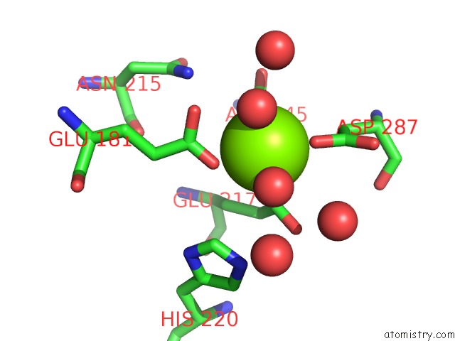
Mono view
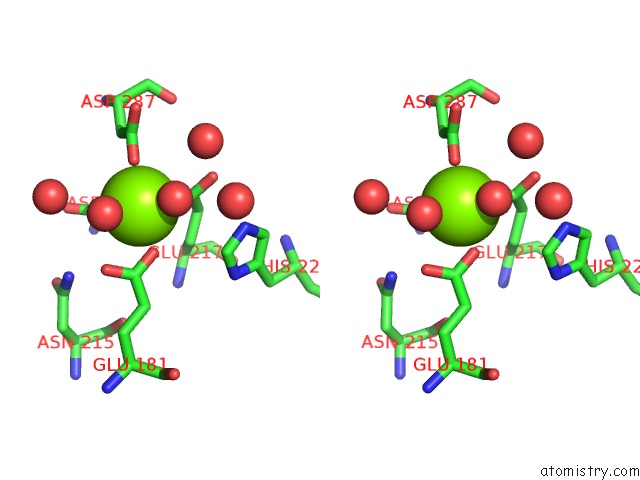
Stereo pair view
Mono view
Stereo pair view
A full contact list of Magnesium with other atoms in the Mg binding
site number 1 of Crystal Structure of Glucose Isomerase Soaked with Glucose within 5.0Å range:
|
Reference:
J.E.Bae,
K.Y.Hwang,
K.H.Nam.
Structural Analysis of Substrate Recognition By Glucose Isomerase in MN2+Binding Mode at M2 Site in S. Rubiginosus Biochem. Biophys. Res. V. 503 770 2018COMMUN..
ISSN: ESSN 1090-2104
PubMed: 29909012
DOI: 10.1016/J.BBRC.2018.06.074
Page generated: Mon Sep 30 18:45:21 2024
ISSN: ESSN 1090-2104
PubMed: 29909012
DOI: 10.1016/J.BBRC.2018.06.074
Last articles
Mg in 1YIOMg in 1YHM
Mg in 1YID
Mg in 1YI1
Mg in 1YI0
Mg in 1YHZ
Mg in 1YHL
Mg in 1YHY
Mg in 1YFR
Mg in 1YHN