Magnesium »
PDB 6emn-6f6w »
6f3h »
Magnesium in PDB 6f3h: Crystal Structure of DSS1 Exoribonuclease Active Site Mutant D477N From Candida Glabrata
Protein crystallography data
The structure of Crystal Structure of DSS1 Exoribonuclease Active Site Mutant D477N From Candida Glabrata, PDB code: 6f3h
was solved by
M.Razew,
E.Nowak,
M.Nowotny,
with X-Ray Crystallography technique. A brief refinement statistics is given in the table below:
| Resolution Low / High (Å) | 29.21 / 2.70 |
| Space group | P 1 |
| Cell size a, b, c (Å), α, β, γ (°) | 73.370, 83.230, 110.370, 106.21, 106.56, 90.93 |
| R / Rfree (%) | 19.6 / 24 |
Magnesium Binding Sites:
The binding sites of Magnesium atom in the Crystal Structure of DSS1 Exoribonuclease Active Site Mutant D477N From Candida Glabrata
(pdb code 6f3h). This binding sites where shown within
5.0 Angstroms radius around Magnesium atom.
In total 2 binding sites of Magnesium where determined in the Crystal Structure of DSS1 Exoribonuclease Active Site Mutant D477N From Candida Glabrata, PDB code: 6f3h:
Jump to Magnesium binding site number: 1; 2;
In total 2 binding sites of Magnesium where determined in the Crystal Structure of DSS1 Exoribonuclease Active Site Mutant D477N From Candida Glabrata, PDB code: 6f3h:
Jump to Magnesium binding site number: 1; 2;
Magnesium binding site 1 out of 2 in 6f3h
Go back to
Magnesium binding site 1 out
of 2 in the Crystal Structure of DSS1 Exoribonuclease Active Site Mutant D477N From Candida Glabrata
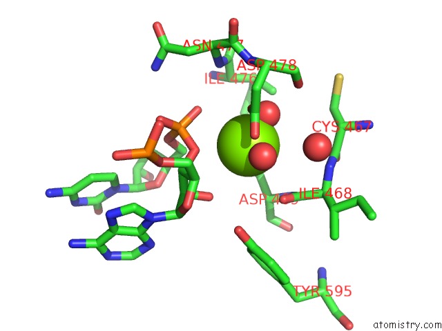
Mono view
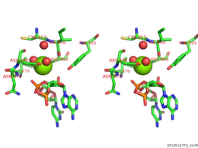
Stereo pair view
Mono view
Stereo pair view
A full contact list of Magnesium with other atoms in the Mg binding
site number 1 of Crystal Structure of DSS1 Exoribonuclease Active Site Mutant D477N From Candida Glabrata within 5.0Å range:
|
Magnesium binding site 2 out of 2 in 6f3h
Go back to
Magnesium binding site 2 out
of 2 in the Crystal Structure of DSS1 Exoribonuclease Active Site Mutant D477N From Candida Glabrata
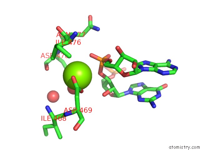
Mono view
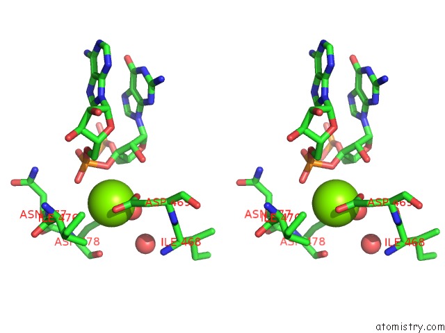
Stereo pair view
Mono view
Stereo pair view
A full contact list of Magnesium with other atoms in the Mg binding
site number 2 of Crystal Structure of DSS1 Exoribonuclease Active Site Mutant D477N From Candida Glabrata within 5.0Å range:
|
Reference:
M.Razew,
Z.Warkocki,
M.Taube,
A.Kolondra,
M.Czarnocki-Cieciura,
E.Nowak,
K.Labedzka-Dmoch,
A.Kawinska,
J.Piatkowski,
P.Golik,
M.Kozak,
A.Dziembowski,
M.Nowotny.
Structural Analysis of Mtexo Mitochondrial Rna Degradosome Reveals Tight Coupling of Nuclease and Helicase Components. Nat Commun V. 9 97 2018.
ISSN: ESSN 2041-1723
PubMed: 29311576
DOI: 10.1038/S41467-017-02570-5
Page generated: Wed Aug 13 05:50:20 2025
ISSN: ESSN 2041-1723
PubMed: 29311576
DOI: 10.1038/S41467-017-02570-5
Last articles
Mg in 6Z5RMg in 6Z5S
Mg in 6Z9J
Mg in 6Z9I
Mg in 6Z9H
Mg in 6Z9A
Mg in 6Z8I
Mg in 6Z8K
Mg in 6Z7T
Mg in 6Z7Z