Magnesium »
PDB 6h5e-6hdi »
6h92 »
Magnesium in PDB 6h92: Phosphorylated Beta-Phosphoglucomutase From Lactococcus Lactis in An Open Conformer to 2.6 A
Enzymatic activity of Phosphorylated Beta-Phosphoglucomutase From Lactococcus Lactis in An Open Conformer to 2.6 A
All present enzymatic activity of Phosphorylated Beta-Phosphoglucomutase From Lactococcus Lactis in An Open Conformer to 2.6 A:
5.4.2.6;
5.4.2.6;
Protein crystallography data
The structure of Phosphorylated Beta-Phosphoglucomutase From Lactococcus Lactis in An Open Conformer to 2.6 A, PDB code: 6h92
was solved by
A.J.Robertson,
C.Bisson,
J.P.Waltho,
with X-Ray Crystallography technique. A brief refinement statistics is given in the table below:
| Resolution Low / High (Å) | 47.82 / 2.60 |
| Space group | P 1 21 1 |
| Cell size a, b, c (Å), α, β, γ (°) | 38.410, 117.180, 52.980, 90.00, 98.66, 90.00 |
| R / Rfree (%) | 23.3 / 31.6 |
Other elements in 6h92:
The structure of Phosphorylated Beta-Phosphoglucomutase From Lactococcus Lactis in An Open Conformer to 2.6 A also contains other interesting chemical elements:
| Sodium | (Na) | 1 atom |
Magnesium Binding Sites:
The binding sites of Magnesium atom in the Phosphorylated Beta-Phosphoglucomutase From Lactococcus Lactis in An Open Conformer to 2.6 A
(pdb code 6h92). This binding sites where shown within
5.0 Angstroms radius around Magnesium atom.
In total only one binding site of Magnesium was determined in the Phosphorylated Beta-Phosphoglucomutase From Lactococcus Lactis in An Open Conformer to 2.6 A, PDB code: 6h92:
In total only one binding site of Magnesium was determined in the Phosphorylated Beta-Phosphoglucomutase From Lactococcus Lactis in An Open Conformer to 2.6 A, PDB code: 6h92:
Magnesium binding site 1 out of 1 in 6h92
Go back to
Magnesium binding site 1 out
of 1 in the Phosphorylated Beta-Phosphoglucomutase From Lactococcus Lactis in An Open Conformer to 2.6 A
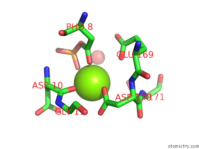
Mono view
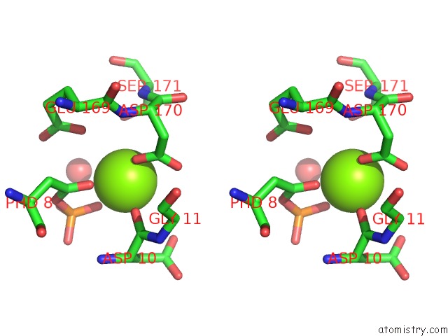
Stereo pair view
Mono view
Stereo pair view
A full contact list of Magnesium with other atoms in the Mg binding
site number 1 of Phosphorylated Beta-Phosphoglucomutase From Lactococcus Lactis in An Open Conformer to 2.6 A within 5.0Å range:
|
Reference:
A.J.Robertson,
C.Bisson,
J.P.Waltho.
Transition State of Phospho-Enzyme Hydrolysis in Beta-Phosphoglucomutase To Be Published.
Page generated: Wed Aug 13 06:54:52 2025
Last articles
Na in 3MSDNa in 3MS8
Na in 3MQQ
Na in 3MQO
Na in 3MQH
Na in 3MQG
Na in 3MP8
Na in 3MPQ
Na in 3MQD
Na in 3MPN