Magnesium »
PDB 6hdh-6hnq »
6hdl »
Magnesium in PDB 6hdl: R49K Variant of Beta-Phosphoglucomutase From Lactococcus Lactis Complexed with Magnesium Trifluoride and Beta-G6P to 1.2 A.
Enzymatic activity of R49K Variant of Beta-Phosphoglucomutase From Lactococcus Lactis Complexed with Magnesium Trifluoride and Beta-G6P to 1.2 A.
All present enzymatic activity of R49K Variant of Beta-Phosphoglucomutase From Lactococcus Lactis Complexed with Magnesium Trifluoride and Beta-G6P to 1.2 A.:
5.4.2.6;
5.4.2.6;
Protein crystallography data
The structure of R49K Variant of Beta-Phosphoglucomutase From Lactococcus Lactis Complexed with Magnesium Trifluoride and Beta-G6P to 1.2 A., PDB code: 6hdl
was solved by
H.P.Wood,
A.J.Robertson,
C.Bisson,
J.P.Waltho,
with X-Ray Crystallography technique. A brief refinement statistics is given in the table below:
| Resolution Low / High (Å) | 35.35 / 1.16 |
| Space group | P 21 21 21 |
| Cell size a, b, c (Å), α, β, γ (°) | 37.550, 54.300, 104.200, 90.00, 90.00, 90.00 |
| R / Rfree (%) | 13.2 / 16.4 |
Other elements in 6hdl:
The structure of R49K Variant of Beta-Phosphoglucomutase From Lactococcus Lactis Complexed with Magnesium Trifluoride and Beta-G6P to 1.2 A. also contains other interesting chemical elements:
| Fluorine | (F) | 3 atoms |
| Sodium | (Na) | 1 atom |
Magnesium Binding Sites:
The binding sites of Magnesium atom in the R49K Variant of Beta-Phosphoglucomutase From Lactococcus Lactis Complexed with Magnesium Trifluoride and Beta-G6P to 1.2 A.
(pdb code 6hdl). This binding sites where shown within
5.0 Angstroms radius around Magnesium atom.
In total 2 binding sites of Magnesium where determined in the R49K Variant of Beta-Phosphoglucomutase From Lactococcus Lactis Complexed with Magnesium Trifluoride and Beta-G6P to 1.2 A., PDB code: 6hdl:
Jump to Magnesium binding site number: 1; 2;
In total 2 binding sites of Magnesium where determined in the R49K Variant of Beta-Phosphoglucomutase From Lactococcus Lactis Complexed with Magnesium Trifluoride and Beta-G6P to 1.2 A., PDB code: 6hdl:
Jump to Magnesium binding site number: 1; 2;
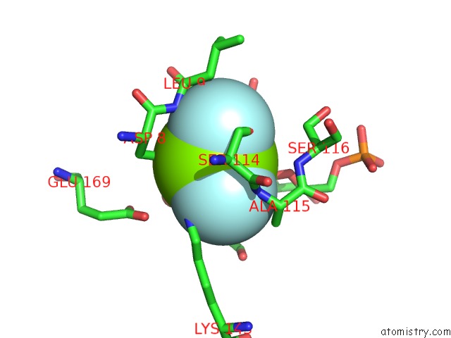
Magnesium binding site 1 out of 2 in 6hdl
Go back to
Magnesium binding site 1 out
of 2 in the R49K Variant of Beta-Phosphoglucomutase From Lactococcus Lactis Complexed with Magnesium Trifluoride and Beta-G6P to 1.2 A.

Mono view
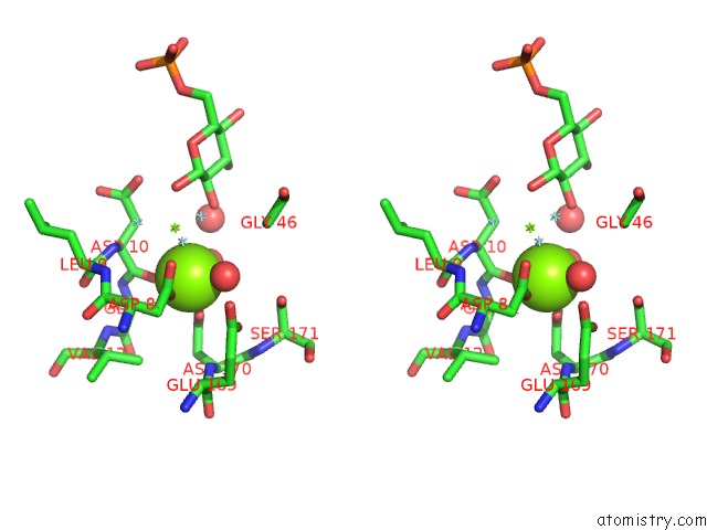
Stereo pair view
Mono view
Stereo pair view
A full contact list of Magnesium with other atoms in the Mg binding
site number 1 of R49K Variant of Beta-Phosphoglucomutase From Lactococcus Lactis Complexed with Magnesium Trifluoride and Beta-G6P to 1.2 A. within 5.0Å range:
|
Magnesium binding site 2 out of 2 in 6hdl
Go back to
Magnesium binding site 2 out
of 2 in the R49K Variant of Beta-Phosphoglucomutase From Lactococcus Lactis Complexed with Magnesium Trifluoride and Beta-G6P to 1.2 A.
Mono view

Stereo pair view
Mono view
Stereo pair view
A full contact list of Magnesium with other atoms in the Mg binding
site number 2 of R49K Variant of Beta-Phosphoglucomutase From Lactococcus Lactis Complexed with Magnesium Trifluoride and Beta-G6P to 1.2 A. within 5.0Å range:
|
Reference:
A.J.Robertson,
C.Bisson,
J.P.Waltho.
Transition State of Phospho-Enzyme Hydrolysis in Beta-Phosphoglucomutase. To Be Published.
Page generated: Tue Oct 1 01:41:59 2024
Last articles
Mg in 5WSAMg in 5WS9
Mg in 5WS8
Mg in 5WS6
Mg in 5WS5
Mg in 5WRT
Mg in 5WRJ
Mg in 5WRI
Mg in 5WQA
Mg in 5WQ0