Magnesium »
PDB 6hns-6hvv »
6hpd »
Magnesium in PDB 6hpd: The Structure of A Beta-Glucuronidase From Glycoside Hydrolase Family 2
Enzymatic activity of The Structure of A Beta-Glucuronidase From Glycoside Hydrolase Family 2
All present enzymatic activity of The Structure of A Beta-Glucuronidase From Glycoside Hydrolase Family 2:
3.2.1.23;
3.2.1.23;
Protein crystallography data
The structure of The Structure of A Beta-Glucuronidase From Glycoside Hydrolase Family 2, PDB code: 6hpd
was solved by
C.S.Robb,
N.Gerlach,
L.Reisky,
U.Bornshoeru,
J.H.Hehemann,
with X-Ray Crystallography technique. A brief refinement statistics is given in the table below:
| Resolution Low / High (Å) | 96.20 / 2.43 |
| Space group | C 1 2 1 |
| Cell size a, b, c (Å), α, β, γ (°) | 157.929, 67.170, 96.720, 90.00, 95.93, 90.00 |
| R / Rfree (%) | 16.5 / 21.1 |
Other elements in 6hpd:
The structure of The Structure of A Beta-Glucuronidase From Glycoside Hydrolase Family 2 also contains other interesting chemical elements:
| Bromine | (Br) | 1 atom |
Magnesium Binding Sites:
The binding sites of Magnesium atom in the The Structure of A Beta-Glucuronidase From Glycoside Hydrolase Family 2
(pdb code 6hpd). This binding sites where shown within
5.0 Angstroms radius around Magnesium atom.
In total only one binding site of Magnesium was determined in the The Structure of A Beta-Glucuronidase From Glycoside Hydrolase Family 2, PDB code: 6hpd:
In total only one binding site of Magnesium was determined in the The Structure of A Beta-Glucuronidase From Glycoside Hydrolase Family 2, PDB code: 6hpd:
Magnesium binding site 1 out of 1 in 6hpd
Go back to
Magnesium binding site 1 out
of 1 in the The Structure of A Beta-Glucuronidase From Glycoside Hydrolase Family 2
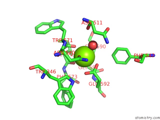
Mono view
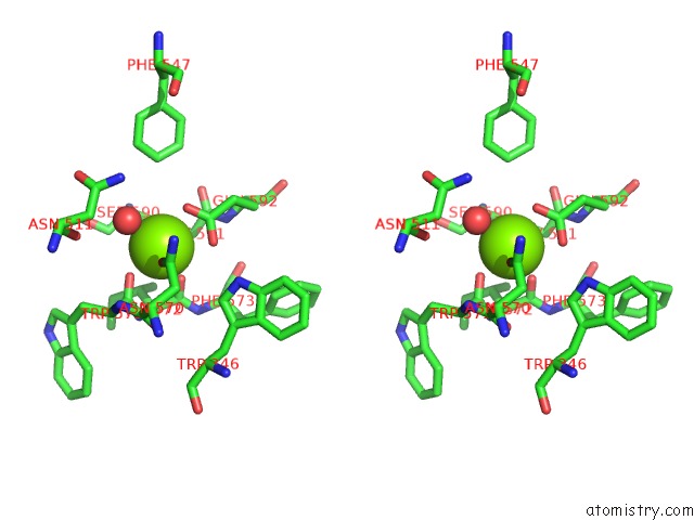
Stereo pair view
Mono view
Stereo pair view
A full contact list of Magnesium with other atoms in the Mg binding
site number 1 of The Structure of A Beta-Glucuronidase From Glycoside Hydrolase Family 2 within 5.0Å range:
|
Reference:
L.Reisky,
A.Prechoux,
M.K.Zuhlke,
M.Baumgen,
C.S.Robb,
N.Gerlach,
T.Roret,
C.Stanetty,
R.Larocque,
G.Michel,
T.Song,
S.Markert,
F.Unfried,
M.D.Mihovilovic,
A.Trautwein-Schult,
D.Becher,
T.Schweder,
U.T.Bornscheuer,
J.H.Hehemann.
A Marine Bacterial Enzymatic Cascade Degrades the Algal Polysaccharide Ulvan. Nat.Chem.Biol. V. 15 803 2019.
ISSN: ESSN 1552-4469
PubMed: 31285597
DOI: 10.1038/S41589-019-0311-9
Page generated: Tue Oct 1 02:11:11 2024
ISSN: ESSN 1552-4469
PubMed: 31285597
DOI: 10.1038/S41589-019-0311-9
Last articles
Fe in 7ZC6Fe in 7ZDL
Fe in 7ZDG
Fe in 7ZCI
Fe in 7ZC5
Fe in 7ZDF
Fe in 7ZDC
Fe in 7ZDB
Fe in 7ZD6
Fe in 7ZCL