Magnesium »
PDB 6iae-6ijj »
6iag »
Magnesium in PDB 6iag: Crystal Structure of Human Phosphodiesterase 4D2 Catalytic Domain with Inhibitor Npd-637
Enzymatic activity of Crystal Structure of Human Phosphodiesterase 4D2 Catalytic Domain with Inhibitor Npd-637
All present enzymatic activity of Crystal Structure of Human Phosphodiesterase 4D2 Catalytic Domain with Inhibitor Npd-637:
3.1.4.53;
3.1.4.53;
Protein crystallography data
The structure of Crystal Structure of Human Phosphodiesterase 4D2 Catalytic Domain with Inhibitor Npd-637, PDB code: 6iag
was solved by
A.K.Singh,
D.G.Brown,
with X-Ray Crystallography technique. A brief refinement statistics is given in the table below:
| Resolution Low / High (Å) | 84.50 / 2.00 |
| Space group | P 21 21 21 |
| Cell size a, b, c (Å), α, β, γ (°) | 99.108, 110.311, 160.722, 90.00, 90.00, 90.00 |
| R / Rfree (%) | 20 / 24.6 |
Other elements in 6iag:
The structure of Crystal Structure of Human Phosphodiesterase 4D2 Catalytic Domain with Inhibitor Npd-637 also contains other interesting chemical elements:
| Zinc | (Zn) | 4 atoms |
Magnesium Binding Sites:
The binding sites of Magnesium atom in the Crystal Structure of Human Phosphodiesterase 4D2 Catalytic Domain with Inhibitor Npd-637
(pdb code 6iag). This binding sites where shown within
5.0 Angstroms radius around Magnesium atom.
In total 4 binding sites of Magnesium where determined in the Crystal Structure of Human Phosphodiesterase 4D2 Catalytic Domain with Inhibitor Npd-637, PDB code: 6iag:
Jump to Magnesium binding site number: 1; 2; 3; 4;
In total 4 binding sites of Magnesium where determined in the Crystal Structure of Human Phosphodiesterase 4D2 Catalytic Domain with Inhibitor Npd-637, PDB code: 6iag:
Jump to Magnesium binding site number: 1; 2; 3; 4;
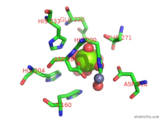
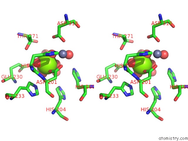
Magnesium binding site 1 out of 4 in 6iag
Go back to
Magnesium binding site 1 out
of 4 in the Crystal Structure of Human Phosphodiesterase 4D2 Catalytic Domain with Inhibitor Npd-637
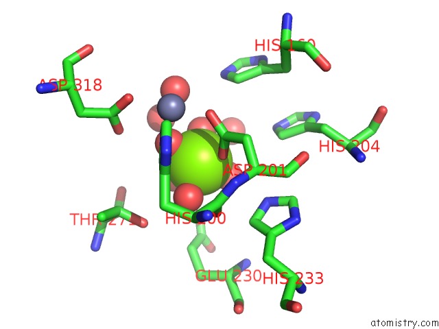
Mono view
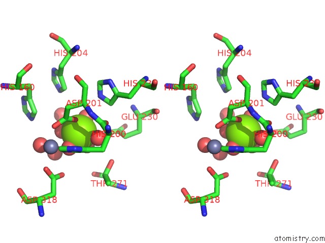
Stereo pair view
Mono view
Stereo pair view
A full contact list of Magnesium with other atoms in the Mg binding
site number 1 of Crystal Structure of Human Phosphodiesterase 4D2 Catalytic Domain with Inhibitor Npd-637 within 5.0Å range:
|
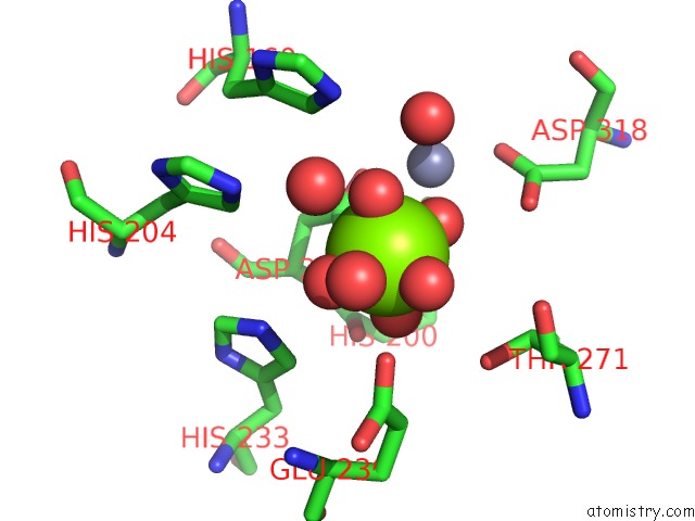
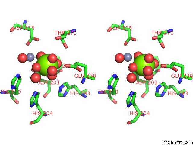
Magnesium binding site 2 out of 4 in 6iag
Go back to
Magnesium binding site 2 out
of 4 in the Crystal Structure of Human Phosphodiesterase 4D2 Catalytic Domain with Inhibitor Npd-637
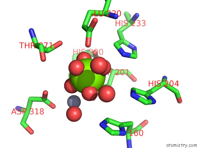
Mono view
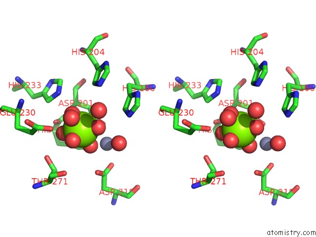
Stereo pair view
Mono view
Stereo pair view
A full contact list of Magnesium with other atoms in the Mg binding
site number 2 of Crystal Structure of Human Phosphodiesterase 4D2 Catalytic Domain with Inhibitor Npd-637 within 5.0Å range:
|
Magnesium binding site 3 out of 4 in 6iag
Go back to
Magnesium binding site 3 out
of 4 in the Crystal Structure of Human Phosphodiesterase 4D2 Catalytic Domain with Inhibitor Npd-637
Mono view
Stereo pair view
Mono view
Stereo pair view
A full contact list of Magnesium with other atoms in the Mg binding
site number 3 of Crystal Structure of Human Phosphodiesterase 4D2 Catalytic Domain with Inhibitor Npd-637 within 5.0Å range:
|
Magnesium binding site 4 out of 4 in 6iag
Go back to
Magnesium binding site 4 out
of 4 in the Crystal Structure of Human Phosphodiesterase 4D2 Catalytic Domain with Inhibitor Npd-637
Mono view
Stereo pair view
Mono view
Stereo pair view
A full contact list of Magnesium with other atoms in the Mg binding
site number 4 of Crystal Structure of Human Phosphodiesterase 4D2 Catalytic Domain with Inhibitor Npd-637 within 5.0Å range:
|
Reference:
A.K.Singh,
D.G.Brown.
HPDE4D2 Structure with Inhibitor Npd-637 To Be Published.
Page generated: Tue Oct 1 03:17:17 2024
Last articles
K in 8CTZK in 8CTX
K in 8CGV
K in 8CTS
K in 8CTT
K in 8CTN
K in 8CGK
K in 8CSU
K in 8CST
K in 8CSS