Magnesium »
PDB 6j7p-6jjw »
6jju »
Magnesium in PDB 6jju: Structure of CA2+ Atpase
Enzymatic activity of Structure of CA2+ Atpase
All present enzymatic activity of Structure of CA2+ Atpase:
7.2.2.10;
7.2.2.10;
Protein crystallography data
The structure of Structure of CA2+ Atpase, PDB code: 6jju
was solved by
M.Inoue,
N.Sakuta,
S.Watanabe,
K.Inaba,
with X-Ray Crystallography technique. A brief refinement statistics is given in the table below:
| Resolution Low / High (Å) | 49.48 / 3.20 |
| Space group | P 21 21 2 |
| Cell size a, b, c (Å), α, β, γ (°) | 165.576, 84.358, 122.187, 90.00, 90.00, 90.00 |
| R / Rfree (%) | 21.1 / 25.6 |
Other elements in 6jju:
The structure of Structure of CA2+ Atpase also contains other interesting chemical elements:
| Calcium | (Ca) | 2 atoms |
Magnesium Binding Sites:
The binding sites of Magnesium atom in the Structure of CA2+ Atpase
(pdb code 6jju). This binding sites where shown within
5.0 Angstroms radius around Magnesium atom.
In total only one binding site of Magnesium was determined in the Structure of CA2+ Atpase, PDB code: 6jju:
In total only one binding site of Magnesium was determined in the Structure of CA2+ Atpase, PDB code: 6jju:
Magnesium binding site 1 out of 1 in 6jju
Go back to
Magnesium binding site 1 out
of 1 in the Structure of CA2+ Atpase
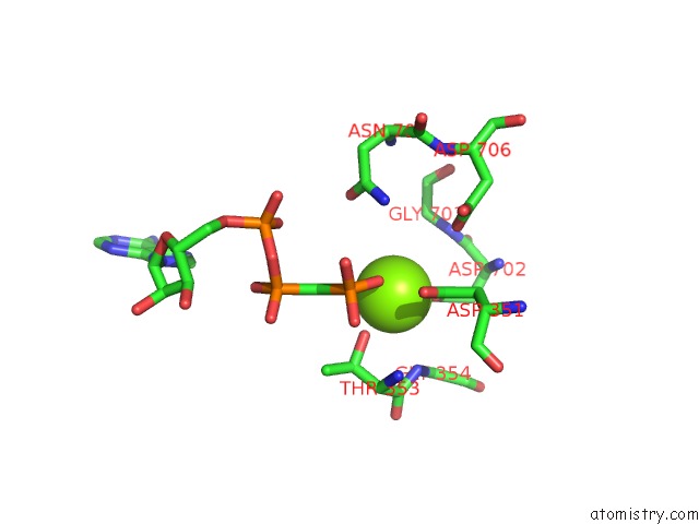
Mono view
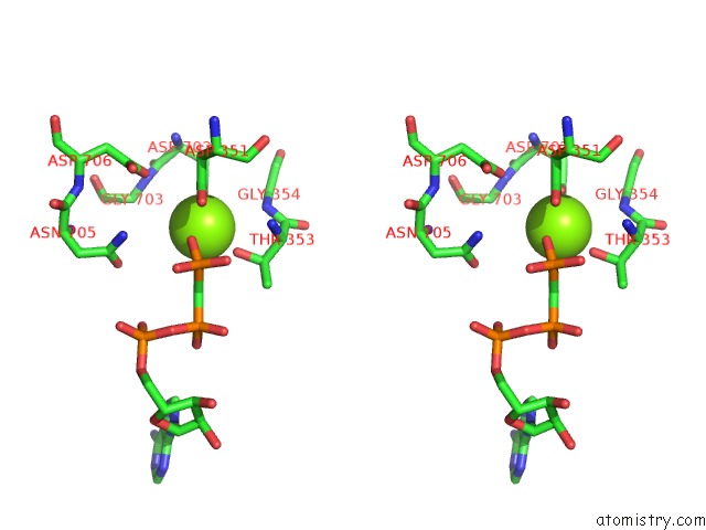
Stereo pair view
Mono view
Stereo pair view
A full contact list of Magnesium with other atoms in the Mg binding
site number 1 of Structure of CA2+ Atpase within 5.0Å range:
|
Reference:
M.Inoue,
N.Sakuta,
S.Watanabe,
Y.Zhang,
K.Yoshikaie,
Y.Tanaka,
R.Ushioda,
Y.Kato,
J.Takagi,
T.Tsukazaki,
K.Nagata,
K.Inaba.
Structural Basis of Sarco/Endoplasmic Reticulum CA2+-Atpase 2B Regulation Via Transmembrane Helix Interplay. Cell Rep V. 27 1221 2019.
ISSN: ESSN 2211-1247
PubMed: 31018135
DOI: 10.1016/J.CELREP.2019.03.106
Page generated: Tue Oct 1 04:59:41 2024
ISSN: ESSN 2211-1247
PubMed: 31018135
DOI: 10.1016/J.CELREP.2019.03.106
Last articles
Fe in 2YXOFe in 2YRS
Fe in 2YXC
Fe in 2YNM
Fe in 2YVJ
Fe in 2YP1
Fe in 2YU2
Fe in 2YU1
Fe in 2YQB
Fe in 2YOO