Magnesium »
PDB 6jwv-6k7k »
6k1e »
Magnesium in PDB 6k1e: Crystal Structure of EXD2 Exonuclease Domain Soaked in Mg and Gmp
Enzymatic activity of Crystal Structure of EXD2 Exonuclease Domain Soaked in Mg and Gmp
All present enzymatic activity of Crystal Structure of EXD2 Exonuclease Domain Soaked in Mg and Gmp:
3.1.11.1;
3.1.11.1;
Protein crystallography data
The structure of Crystal Structure of EXD2 Exonuclease Domain Soaked in Mg and Gmp, PDB code: 6k1e
was solved by
J.Park,
C.Lee,
with X-Ray Crystallography technique. A brief refinement statistics is given in the table below:
| Resolution Low / High (Å) | 49.47 / 2.90 |
| Space group | P 21 21 21 |
| Cell size a, b, c (Å), α, β, γ (°) | 46.496, 73.288, 134.086, 90.00, 90.00, 90.00 |
| R / Rfree (%) | 22.8 / 26.8 |
Magnesium Binding Sites:
The binding sites of Magnesium atom in the Crystal Structure of EXD2 Exonuclease Domain Soaked in Mg and Gmp
(pdb code 6k1e). This binding sites where shown within
5.0 Angstroms radius around Magnesium atom.
In total 4 binding sites of Magnesium where determined in the Crystal Structure of EXD2 Exonuclease Domain Soaked in Mg and Gmp, PDB code: 6k1e:
Jump to Magnesium binding site number: 1; 2; 3; 4;
In total 4 binding sites of Magnesium where determined in the Crystal Structure of EXD2 Exonuclease Domain Soaked in Mg and Gmp, PDB code: 6k1e:
Jump to Magnesium binding site number: 1; 2; 3; 4;
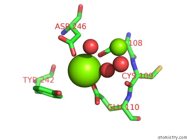
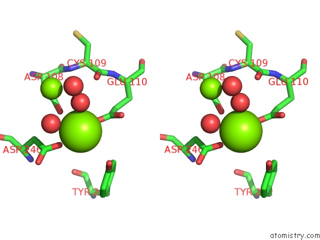
Magnesium binding site 1 out of 4 in 6k1e
Go back to
Magnesium binding site 1 out
of 4 in the Crystal Structure of EXD2 Exonuclease Domain Soaked in Mg and Gmp
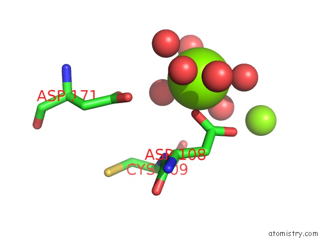
Mono view
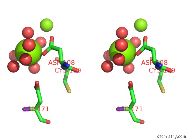
Stereo pair view
Mono view
Stereo pair view
A full contact list of Magnesium with other atoms in the Mg binding
site number 1 of Crystal Structure of EXD2 Exonuclease Domain Soaked in Mg and Gmp within 5.0Å range:
|
Magnesium binding site 2 out of 4 in 6k1e
Go back to
Magnesium binding site 2 out
of 4 in the Crystal Structure of EXD2 Exonuclease Domain Soaked in Mg and Gmp
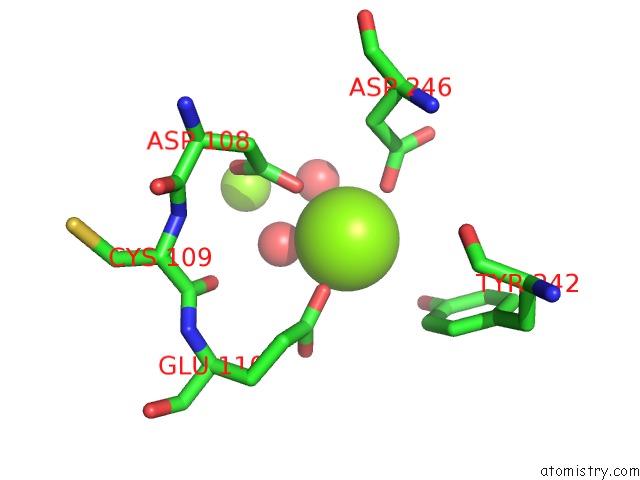
Mono view
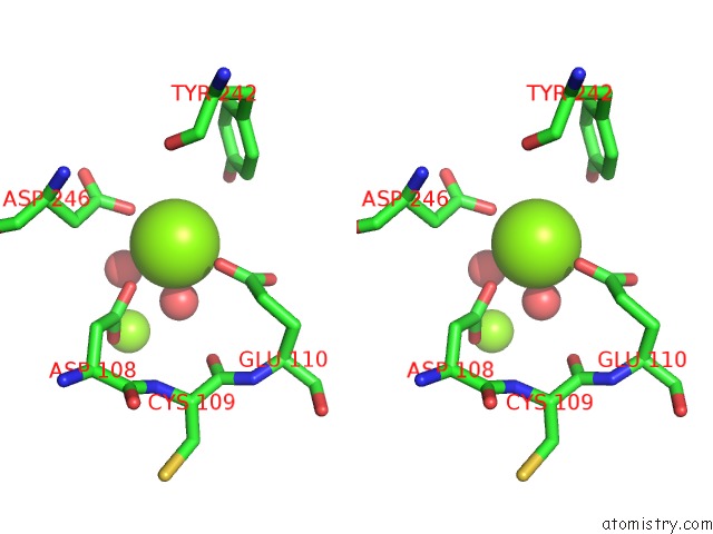
Stereo pair view
Mono view
Stereo pair view
A full contact list of Magnesium with other atoms in the Mg binding
site number 2 of Crystal Structure of EXD2 Exonuclease Domain Soaked in Mg and Gmp within 5.0Å range:
|
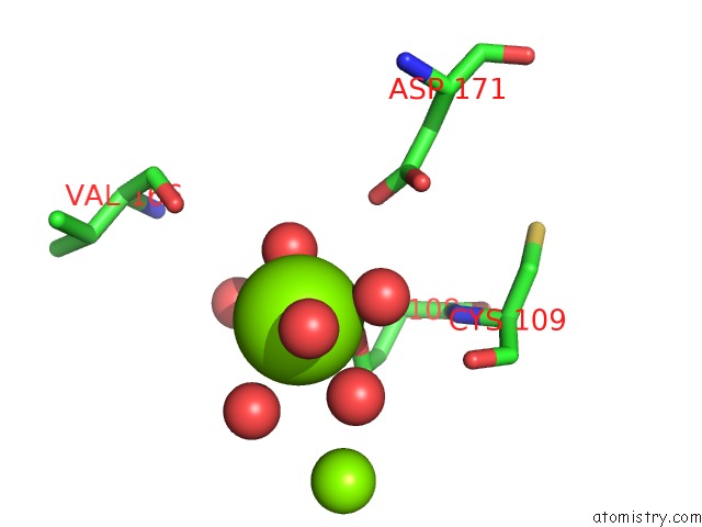
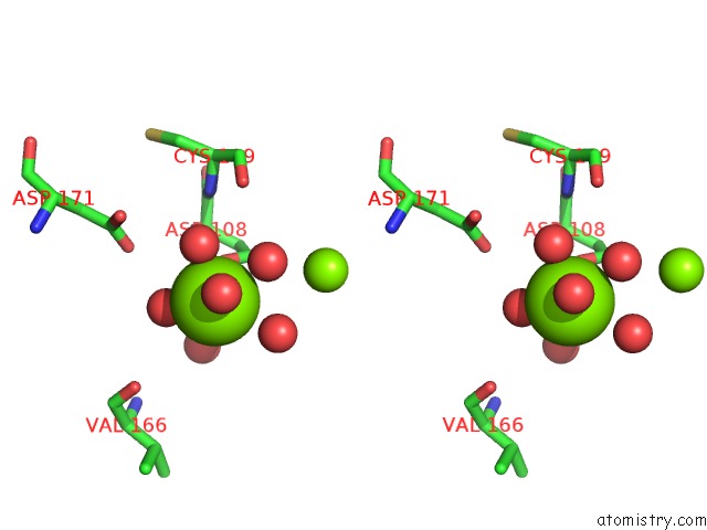
Magnesium binding site 3 out of 4 in 6k1e
Go back to
Magnesium binding site 3 out
of 4 in the Crystal Structure of EXD2 Exonuclease Domain Soaked in Mg and Gmp
Mono view
Stereo pair view
Mono view
Stereo pair view
A full contact list of Magnesium with other atoms in the Mg binding
site number 3 of Crystal Structure of EXD2 Exonuclease Domain Soaked in Mg and Gmp within 5.0Å range:
|
Magnesium binding site 4 out of 4 in 6k1e
Go back to
Magnesium binding site 4 out
of 4 in the Crystal Structure of EXD2 Exonuclease Domain Soaked in Mg and Gmp
Mono view
Stereo pair view
Mono view
Stereo pair view
A full contact list of Magnesium with other atoms in the Mg binding
site number 4 of Crystal Structure of EXD2 Exonuclease Domain Soaked in Mg and Gmp within 5.0Å range:
|
Reference:
J.Park,
S.Y.Lee,
H.Jeong,
M.G.Kang,
L.Van Haute,
M.Minczuk,
J.K.Seo,
Y.Jun,
K.Myung,
H.W.Rhee,
C.Lee.
The Structure of Human EXD2 Reveals A Chimeric 3' to 5' Exonuclease Domain That Discriminates Substrates Via Metal Coordination. Nucleic Acids Res. V. 47 7078 2019.
ISSN: ESSN 1362-4962
PubMed: 31127291
DOI: 10.1093/NAR/GKZ454
Page generated: Wed Aug 13 10:11:25 2025
ISSN: ESSN 1362-4962
PubMed: 31127291
DOI: 10.1093/NAR/GKZ454
Last articles
Mg in 6LU1Mg in 6LT4
Mg in 6LY7
Mg in 6LY6
Mg in 6LY3
Mg in 6LX1
Mg in 6LTW
Mg in 6LVW
Mg in 6LUH
Mg in 6LTS