Magnesium »
PDB 6n7n-6nk8 »
6nb2 »
Magnesium in PDB 6nb2: Crystal Structure of Enolase From Legionella Pneumophila Bound to 2- Phosphoglyceric Acid and Magnesium
Enzymatic activity of Crystal Structure of Enolase From Legionella Pneumophila Bound to 2- Phosphoglyceric Acid and Magnesium
All present enzymatic activity of Crystal Structure of Enolase From Legionella Pneumophila Bound to 2- Phosphoglyceric Acid and Magnesium:
4.2.1.11;
4.2.1.11;
Protein crystallography data
The structure of Crystal Structure of Enolase From Legionella Pneumophila Bound to 2- Phosphoglyceric Acid and Magnesium, PDB code: 6nb2
was solved by
Seattle Structural Genomics Center For Infectious Disease (Ssgcid),
with X-Ray Crystallography technique. A brief refinement statistics is given in the table below:
| Resolution Low / High (Å) | 45.12 / 1.85 |
| Space group | P 42 21 2 |
| Cell size a, b, c (Å), α, β, γ (°) | 116.450, 116.450, 142.780, 90.00, 90.00, 90.00 |
| R / Rfree (%) | 14.6 / 17.2 |
Magnesium Binding Sites:
The binding sites of Magnesium atom in the Crystal Structure of Enolase From Legionella Pneumophila Bound to 2- Phosphoglyceric Acid and Magnesium
(pdb code 6nb2). This binding sites where shown within
5.0 Angstroms radius around Magnesium atom.
In total 4 binding sites of Magnesium where determined in the Crystal Structure of Enolase From Legionella Pneumophila Bound to 2- Phosphoglyceric Acid and Magnesium, PDB code: 6nb2:
Jump to Magnesium binding site number: 1; 2; 3; 4;
In total 4 binding sites of Magnesium where determined in the Crystal Structure of Enolase From Legionella Pneumophila Bound to 2- Phosphoglyceric Acid and Magnesium, PDB code: 6nb2:
Jump to Magnesium binding site number: 1; 2; 3; 4;
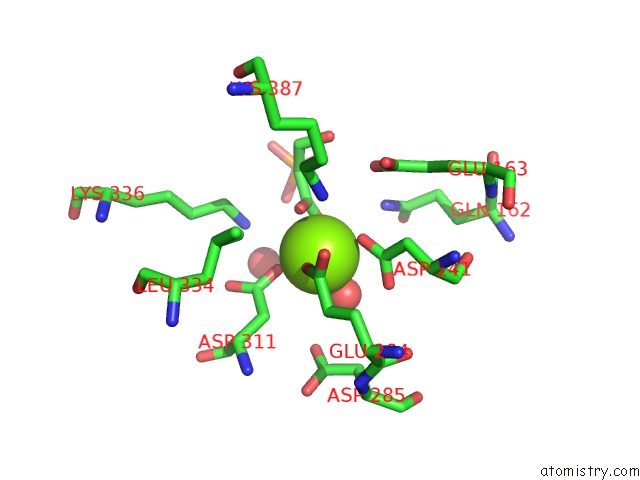
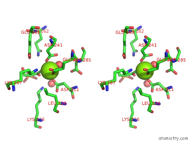
Magnesium binding site 1 out of 4 in 6nb2
Go back to
Magnesium binding site 1 out
of 4 in the Crystal Structure of Enolase From Legionella Pneumophila Bound to 2- Phosphoglyceric Acid and Magnesium
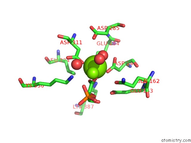
Mono view
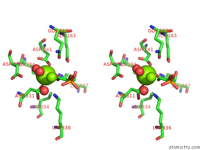
Stereo pair view
Mono view
Stereo pair view
A full contact list of Magnesium with other atoms in the Mg binding
site number 1 of Crystal Structure of Enolase From Legionella Pneumophila Bound to 2- Phosphoglyceric Acid and Magnesium within 5.0Å range:
|
Magnesium binding site 2 out of 4 in 6nb2
Go back to
Magnesium binding site 2 out
of 4 in the Crystal Structure of Enolase From Legionella Pneumophila Bound to 2- Phosphoglyceric Acid and Magnesium
Mono view
Stereo pair view
Mono view
Stereo pair view
A full contact list of Magnesium with other atoms in the Mg binding
site number 2 of Crystal Structure of Enolase From Legionella Pneumophila Bound to 2- Phosphoglyceric Acid and Magnesium within 5.0Å range:
|
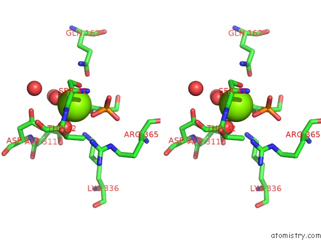
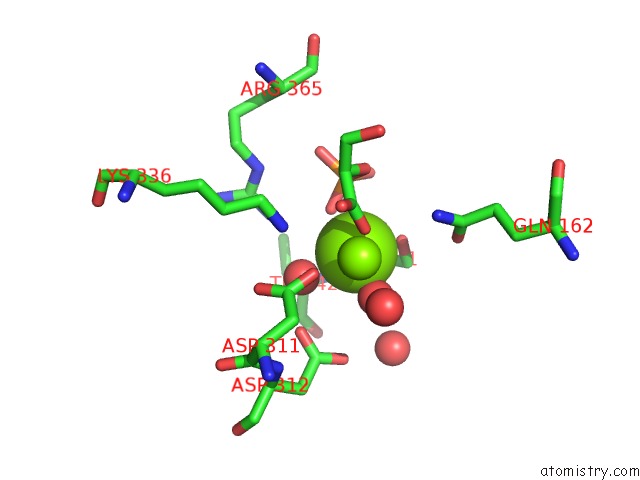
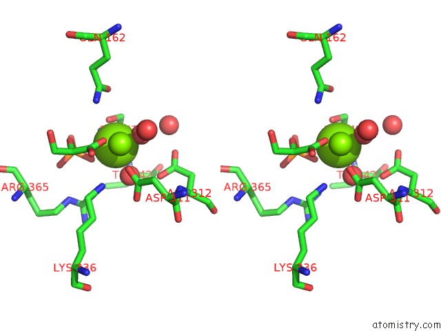
Magnesium binding site 3 out of 4 in 6nb2
Go back to
Magnesium binding site 3 out
of 4 in the Crystal Structure of Enolase From Legionella Pneumophila Bound to 2- Phosphoglyceric Acid and Magnesium
Mono view
Stereo pair view
Mono view
Stereo pair view
A full contact list of Magnesium with other atoms in the Mg binding
site number 3 of Crystal Structure of Enolase From Legionella Pneumophila Bound to 2- Phosphoglyceric Acid and Magnesium within 5.0Å range:
|
Magnesium binding site 4 out of 4 in 6nb2
Go back to
Magnesium binding site 4 out
of 4 in the Crystal Structure of Enolase From Legionella Pneumophila Bound to 2- Phosphoglyceric Acid and Magnesium
Mono view
Stereo pair view
Mono view
Stereo pair view
A full contact list of Magnesium with other atoms in the Mg binding
site number 4 of Crystal Structure of Enolase From Legionella Pneumophila Bound to 2- Phosphoglyceric Acid and Magnesium within 5.0Å range:
|
Reference:
D.R.Davies,
J.Abendroth,
D.D.Lorimer,
T.E.Edwards.
Crystal Structure of Enolase From Legionella Pneumophila Bound to 2-Phosphoglyceric Acid and Magnesium To Be Published.
Page generated: Tue Oct 1 12:37:27 2024
Last articles
I in 4RCFI in 4R11
I in 4RCE
I in 4RCD
I in 4R1T
I in 4QRP
I in 4QU3
I in 4PXS
I in 4QJQ
I in 4Q53