Magnesium »
PDB 6n7n-6nk8 »
6njd »
Magnesium in PDB 6njd: Crystal Structure of Ravd (Residues 1-200) From Legionella Pneumophila (Strain Corby) Complexed with Met-1 Linked Di-Ubiquitin
Protein crystallography data
The structure of Crystal Structure of Ravd (Residues 1-200) From Legionella Pneumophila (Strain Corby) Complexed with Met-1 Linked Di-Ubiquitin, PDB code: 6njd
was solved by
X.Wang,
Y.Zhou,
Y.Zhu,
with X-Ray Crystallography technique. A brief refinement statistics is given in the table below:
| Resolution Low / High (Å) | 50.01 / 2.05 |
| Space group | P 21 21 2 |
| Cell size a, b, c (Å), α, β, γ (°) | 77.740, 102.090, 90.920, 90.00, 90.00, 90.00 |
| R / Rfree (%) | 21.2 / 24.6 |
Magnesium Binding Sites:
The binding sites of Magnesium atom in the Crystal Structure of Ravd (Residues 1-200) From Legionella Pneumophila (Strain Corby) Complexed with Met-1 Linked Di-Ubiquitin
(pdb code 6njd). This binding sites where shown within
5.0 Angstroms radius around Magnesium atom.
In total 2 binding sites of Magnesium where determined in the Crystal Structure of Ravd (Residues 1-200) From Legionella Pneumophila (Strain Corby) Complexed with Met-1 Linked Di-Ubiquitin, PDB code: 6njd:
Jump to Magnesium binding site number: 1; 2;
In total 2 binding sites of Magnesium where determined in the Crystal Structure of Ravd (Residues 1-200) From Legionella Pneumophila (Strain Corby) Complexed with Met-1 Linked Di-Ubiquitin, PDB code: 6njd:
Jump to Magnesium binding site number: 1; 2;
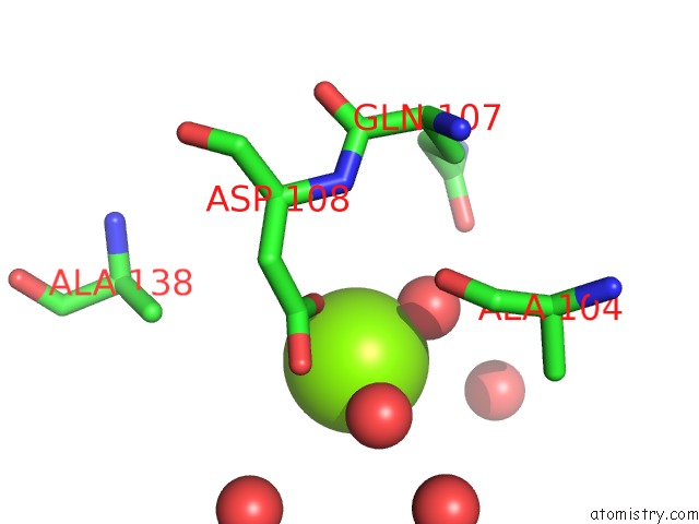
Magnesium binding site 1 out of 2 in 6njd
Go back to
Magnesium binding site 1 out
of 2 in the Crystal Structure of Ravd (Residues 1-200) From Legionella Pneumophila (Strain Corby) Complexed with Met-1 Linked Di-Ubiquitin
Mono view

Stereo pair view
Mono view
Stereo pair view
A full contact list of Magnesium with other atoms in the Mg binding
site number 1 of Crystal Structure of Ravd (Residues 1-200) From Legionella Pneumophila (Strain Corby) Complexed with Met-1 Linked Di-Ubiquitin within 5.0Å range:
|
Magnesium binding site 2 out of 2 in 6njd
Go back to
Magnesium binding site 2 out
of 2 in the Crystal Structure of Ravd (Residues 1-200) From Legionella Pneumophila (Strain Corby) Complexed with Met-1 Linked Di-Ubiquitin

Mono view

Stereo pair view
Mono view
Stereo pair view
A full contact list of Magnesium with other atoms in the Mg binding
site number 2 of Crystal Structure of Ravd (Residues 1-200) From Legionella Pneumophila (Strain Corby) Complexed with Met-1 Linked Di-Ubiquitin within 5.0Å range:
|
Reference:
M.Wan,
X.Wang,
C.Huang,
D.Xu,
Z.Wang,
Y.Zhou,
Y.Zhu.
A Bacterial Effector Deubiquitinase Specifically Hydrolyses Linear Ubiquitin Chains to Inhibit Host Inflammatory Signalling. Nat Microbiol V. 4 1282 2019.
ISSN: ESSN 2058-5276
PubMed: 31110362
DOI: 10.1038/S41564-019-0454-1
Page generated: Tue Oct 1 12:40:24 2024
ISSN: ESSN 2058-5276
PubMed: 31110362
DOI: 10.1038/S41564-019-0454-1
Last articles
I in 4S22I in 4S2H
I in 4QX5
I in 4S2G
I in 4S2F
I in 4RX1
I in 4S2D
I in 4RYM
I in 4REC
I in 4RCF