Magnesium »
PDB 6nop-6o2r »
6ny6 »
Magnesium in PDB 6ny6: Structure of Dimeric Escherichia Coli Toxin Yoeb Bound to the Thermus Thermophilus 30S Ribosome
Protein crystallography data
The structure of Structure of Dimeric Escherichia Coli Toxin Yoeb Bound to the Thermus Thermophilus 30S Ribosome, PDB code: 6ny6
was solved by
I.J.Pavelich,
E.D.Hoffer,
T.Maehigashi,
C.M.Dunham,
with X-Ray Crystallography technique. A brief refinement statistics is given in the table below:
| Resolution Low / High (Å) | 49.83 / 3.74 |
| Space group | P 41 21 2 |
| Cell size a, b, c (Å), α, β, γ (°) | 401.710, 401.710, 175.440, 90.00, 90.00, 90.00 |
| R / Rfree (%) | 20.6 / 22.4 |
Other elements in 6ny6:
The structure of Structure of Dimeric Escherichia Coli Toxin Yoeb Bound to the Thermus Thermophilus 30S Ribosome also contains other interesting chemical elements:
| Zinc | (Zn) | 1 atom |
| Iron | (Fe) | 4 atoms |
Magnesium Binding Sites:
Pages:
>>> Page 1 <<< Page 2, Binding sites: 11 - 20; Page 3, Binding sites: 21 - 30; Page 4, Binding sites: 31 - 40; Page 5, Binding sites: 41 - 50; Page 6, Binding sites: 51 - 60; Page 7, Binding sites: 61 - 70; Page 8, Binding sites: 71 - 80; Page 9, Binding sites: 81 - 90; Page 10, Binding sites: 91 - 100; Page 11, Binding sites: 101 - 110; Page 12, Binding sites: 111 - 120; Page 13, Binding sites: 121 - 130; Page 14, Binding sites: 131 - 140; Page 15, Binding sites: 141 - 150; Page 16, Binding sites: 151 - 160; Page 17, Binding sites: 161 - 170; Page 18, Binding sites: 171 - 180; Page 19, Binding sites: 181 - 184;Binding sites:
The binding sites of Magnesium atom in the Structure of Dimeric Escherichia Coli Toxin Yoeb Bound to the Thermus Thermophilus 30S Ribosome (pdb code 6ny6). This binding sites where shown within 5.0 Angstroms radius around Magnesium atom.In total 184 binding sites of Magnesium where determined in the Structure of Dimeric Escherichia Coli Toxin Yoeb Bound to the Thermus Thermophilus 30S Ribosome, PDB code: 6ny6:
Jump to Magnesium binding site number: 1; 2; 3; 4; 5; 6; 7; 8; 9; 10;
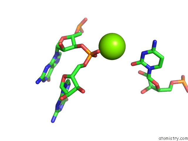
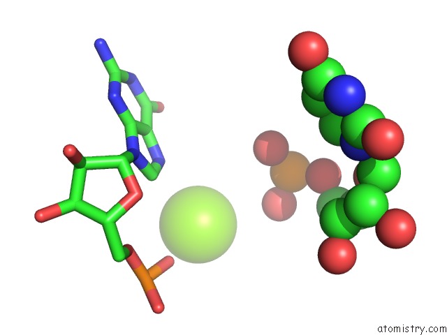
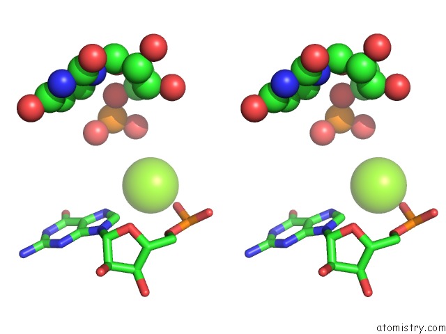
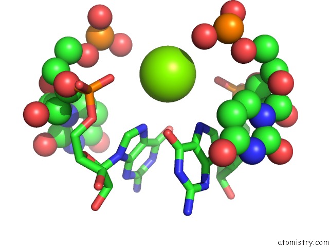
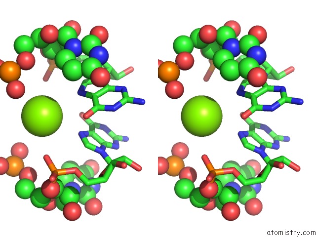
Magnesium binding site 1 out of 184 in 6ny6
Go back to
Magnesium binding site 1 out
of 184 in the Structure of Dimeric Escherichia Coli Toxin Yoeb Bound to the Thermus Thermophilus 30S Ribosome
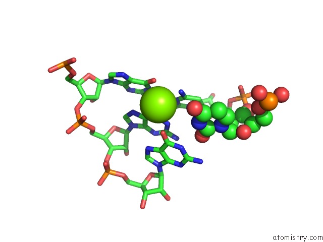
Mono view
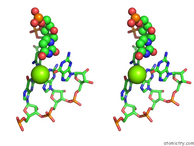
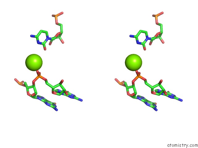
Stereo pair view
Mono view
Stereo pair view
A full contact list of Magnesium with other atoms in the Mg binding
site number 1 of Structure of Dimeric Escherichia Coli Toxin Yoeb Bound to the Thermus Thermophilus 30S Ribosome within 5.0Å range:
|
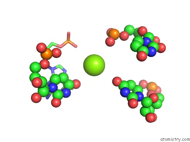
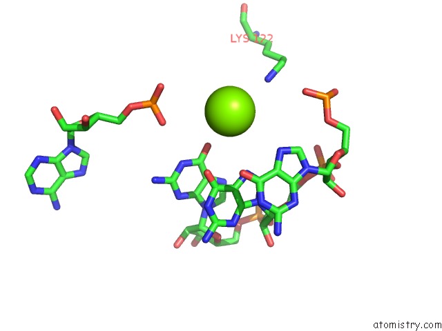
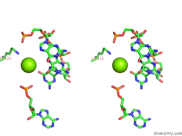
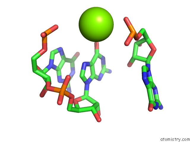
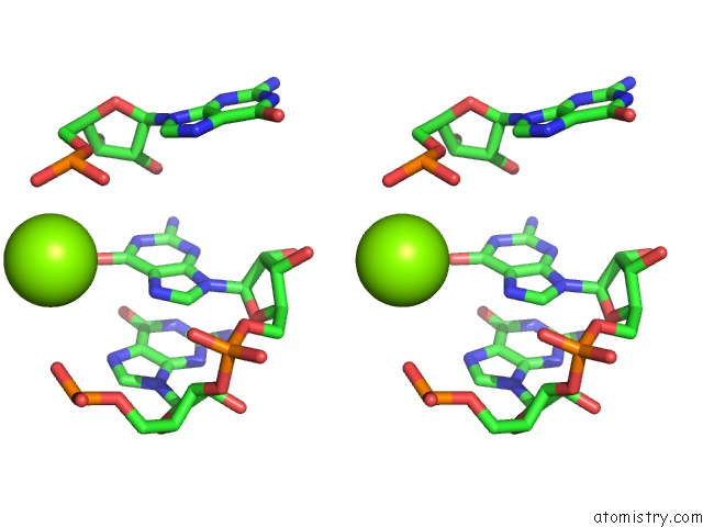
Magnesium binding site 2 out of 184 in 6ny6
Go back to
Magnesium binding site 2 out
of 184 in the Structure of Dimeric Escherichia Coli Toxin Yoeb Bound to the Thermus Thermophilus 30S Ribosome
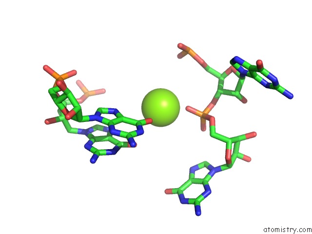
Mono view
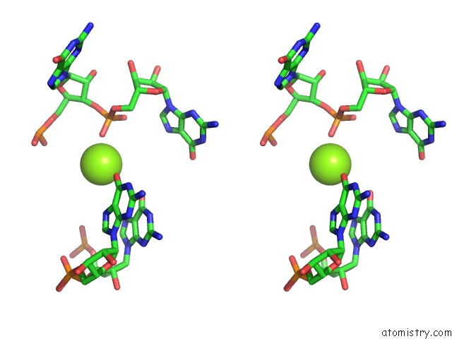
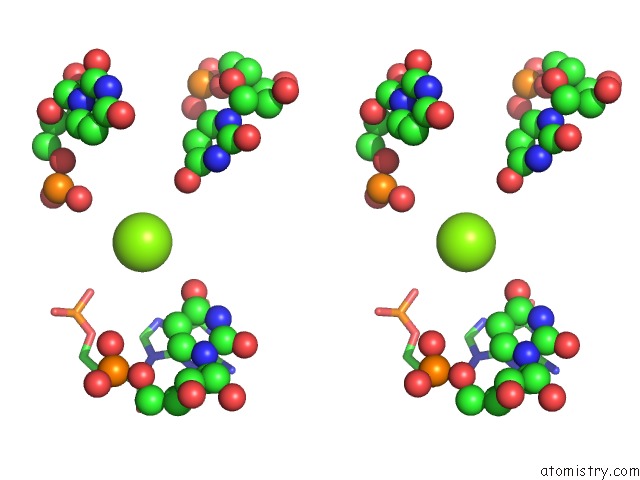
Stereo pair view
Mono view
Stereo pair view
A full contact list of Magnesium with other atoms in the Mg binding
site number 2 of Structure of Dimeric Escherichia Coli Toxin Yoeb Bound to the Thermus Thermophilus 30S Ribosome within 5.0Å range:
|
Magnesium binding site 3 out of 184 in 6ny6
Go back to
Magnesium binding site 3 out
of 184 in the Structure of Dimeric Escherichia Coli Toxin Yoeb Bound to the Thermus Thermophilus 30S Ribosome
Mono view
Stereo pair view
Mono view
Stereo pair view
A full contact list of Magnesium with other atoms in the Mg binding
site number 3 of Structure of Dimeric Escherichia Coli Toxin Yoeb Bound to the Thermus Thermophilus 30S Ribosome within 5.0Å range:
|
Magnesium binding site 4 out of 184 in 6ny6
Go back to
Magnesium binding site 4 out
of 184 in the Structure of Dimeric Escherichia Coli Toxin Yoeb Bound to the Thermus Thermophilus 30S Ribosome
Mono view
Stereo pair view
Mono view
Stereo pair view
A full contact list of Magnesium with other atoms in the Mg binding
site number 4 of Structure of Dimeric Escherichia Coli Toxin Yoeb Bound to the Thermus Thermophilus 30S Ribosome within 5.0Å range:
|
Magnesium binding site 5 out of 184 in 6ny6
Go back to
Magnesium binding site 5 out
of 184 in the Structure of Dimeric Escherichia Coli Toxin Yoeb Bound to the Thermus Thermophilus 30S Ribosome
Mono view
Stereo pair view
Mono view
Stereo pair view
A full contact list of Magnesium with other atoms in the Mg binding
site number 5 of Structure of Dimeric Escherichia Coli Toxin Yoeb Bound to the Thermus Thermophilus 30S Ribosome within 5.0Å range:
|
Magnesium binding site 6 out of 184 in 6ny6
Go back to
Magnesium binding site 6 out
of 184 in the Structure of Dimeric Escherichia Coli Toxin Yoeb Bound to the Thermus Thermophilus 30S Ribosome
Mono view
Stereo pair view
Mono view
Stereo pair view
A full contact list of Magnesium with other atoms in the Mg binding
site number 6 of Structure of Dimeric Escherichia Coli Toxin Yoeb Bound to the Thermus Thermophilus 30S Ribosome within 5.0Å range:
|
Magnesium binding site 7 out of 184 in 6ny6
Go back to
Magnesium binding site 7 out
of 184 in the Structure of Dimeric Escherichia Coli Toxin Yoeb Bound to the Thermus Thermophilus 30S Ribosome
Mono view
Stereo pair view
Mono view
Stereo pair view
A full contact list of Magnesium with other atoms in the Mg binding
site number 7 of Structure of Dimeric Escherichia Coli Toxin Yoeb Bound to the Thermus Thermophilus 30S Ribosome within 5.0Å range:
|
Magnesium binding site 8 out of 184 in 6ny6
Go back to
Magnesium binding site 8 out
of 184 in the Structure of Dimeric Escherichia Coli Toxin Yoeb Bound to the Thermus Thermophilus 30S Ribosome
Mono view
Stereo pair view
Mono view
Stereo pair view
A full contact list of Magnesium with other atoms in the Mg binding
site number 8 of Structure of Dimeric Escherichia Coli Toxin Yoeb Bound to the Thermus Thermophilus 30S Ribosome within 5.0Å range:
|
Magnesium binding site 9 out of 184 in 6ny6
Go back to
Magnesium binding site 9 out
of 184 in the Structure of Dimeric Escherichia Coli Toxin Yoeb Bound to the Thermus Thermophilus 30S Ribosome
Mono view
Stereo pair view
Mono view
Stereo pair view
A full contact list of Magnesium with other atoms in the Mg binding
site number 9 of Structure of Dimeric Escherichia Coli Toxin Yoeb Bound to the Thermus Thermophilus 30S Ribosome within 5.0Å range:
|
Magnesium binding site 10 out of 184 in 6ny6
Go back to
Magnesium binding site 10 out
of 184 in the Structure of Dimeric Escherichia Coli Toxin Yoeb Bound to the Thermus Thermophilus 30S Ribosome
Mono view
Stereo pair view
Mono view
Stereo pair view
A full contact list of Magnesium with other atoms in the Mg binding
site number 10 of Structure of Dimeric Escherichia Coli Toxin Yoeb Bound to the Thermus Thermophilus 30S Ribosome within 5.0Å range:
|
Reference:
I.J.Pavelich,
T.Maehigashi,
E.D.Hoffer,
A.Ruangprasert,
S.J.Miles,
C.M.Dunham.
Monomeric Yoeb Toxin Retains Rnase Activity But Adopts An Obligate Dimeric Form For Thermal Stability. Nucleic Acids Res. V. 47 10400 2019.
ISSN: ESSN 1362-4962
PubMed: 31501867
DOI: 10.1093/NAR/GKZ760
Page generated: Tue Oct 1 12:56:18 2024
ISSN: ESSN 1362-4962
PubMed: 31501867
DOI: 10.1093/NAR/GKZ760
Last articles
Fe in 7V2KFe in 7V2H
Fe in 7V2X
Fe in 7V2U
Fe in 7V2T
Fe in 7V2F
Fe in 7V2E
Fe in 7UUS
Fe in 7V2D
Fe in 7V2C