Magnesium »
PDB 6rl4-6rvm »
6rsw »
Magnesium in PDB 6rsw: Hfd Domain of Mouse CAP1 Bound to the Pointed End of G-Actin
Protein crystallography data
The structure of Hfd Domain of Mouse CAP1 Bound to the Pointed End of G-Actin, PDB code: 6rsw
was solved by
T.Kotila,
K.Kogan,
P.Lappalainen,
with X-Ray Crystallography technique. A brief refinement statistics is given in the table below:
| Resolution Low / High (Å) | 41.45 / 1.95 |
| Space group | P 1 21 1 |
| Cell size a, b, c (Å), α, β, γ (°) | 87.370, 54.490, 87.830, 90.00, 93.61, 90.00 |
| R / Rfree (%) | 16.6 / 19.4 |
Magnesium Binding Sites:
The binding sites of Magnesium atom in the Hfd Domain of Mouse CAP1 Bound to the Pointed End of G-Actin
(pdb code 6rsw). This binding sites where shown within
5.0 Angstroms radius around Magnesium atom.
In total 2 binding sites of Magnesium where determined in the Hfd Domain of Mouse CAP1 Bound to the Pointed End of G-Actin, PDB code: 6rsw:
Jump to Magnesium binding site number: 1; 2;
In total 2 binding sites of Magnesium where determined in the Hfd Domain of Mouse CAP1 Bound to the Pointed End of G-Actin, PDB code: 6rsw:
Jump to Magnesium binding site number: 1; 2;
Magnesium binding site 1 out of 2 in 6rsw
Go back to
Magnesium binding site 1 out
of 2 in the Hfd Domain of Mouse CAP1 Bound to the Pointed End of G-Actin
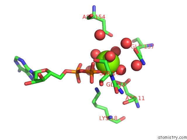
Mono view
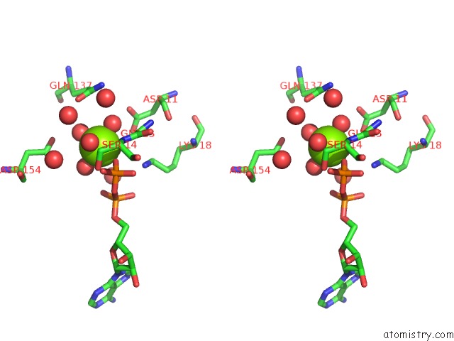
Stereo pair view
Mono view
Stereo pair view
A full contact list of Magnesium with other atoms in the Mg binding
site number 1 of Hfd Domain of Mouse CAP1 Bound to the Pointed End of G-Actin within 5.0Å range:
|
Magnesium binding site 2 out of 2 in 6rsw
Go back to
Magnesium binding site 2 out
of 2 in the Hfd Domain of Mouse CAP1 Bound to the Pointed End of G-Actin
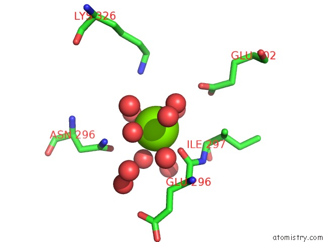
Mono view
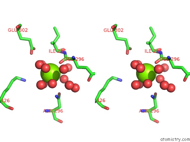
Stereo pair view
Mono view
Stereo pair view
A full contact list of Magnesium with other atoms in the Mg binding
site number 2 of Hfd Domain of Mouse CAP1 Bound to the Pointed End of G-Actin within 5.0Å range:
|
Reference:
T.Kotila,
H.Wioland,
G.Enkavi,
K.Kogan,
I.Vattulainen,
A.Jegou,
G.Romet-Lemonne,
P.Lappalainen.
Mechanism of Synergistic Actin Filament Pointed End Depolymerization By Cyclase-Associated Protein and Cofilin. Nat Commun V. 10 5320 2019.
ISSN: ESSN 2041-1723
PubMed: 31757941
DOI: 10.1038/S41467-019-13213-2
Page generated: Tue Oct 1 17:31:12 2024
ISSN: ESSN 2041-1723
PubMed: 31757941
DOI: 10.1038/S41467-019-13213-2
Last articles
I in 4DZLI in 4E91
I in 4DZN
I in 4DZM
I in 4DGH
I in 4DUS
I in 4DHG
I in 4DH6
I in 4DNY
I in 4CB6