Magnesium »
PDB 6sht-6stf »
6sht »
Magnesium in PDB 6sht: Molecular Structure of Mouse Apoferritin Resolved at 2.7 Angstroms with the Glacios Cryo-Microscope
Enzymatic activity of Molecular Structure of Mouse Apoferritin Resolved at 2.7 Angstroms with the Glacios Cryo-Microscope
All present enzymatic activity of Molecular Structure of Mouse Apoferritin Resolved at 2.7 Angstroms with the Glacios Cryo-Microscope:
1.16.3.1;
1.16.3.1;
Other elements in 6sht:
The structure of Molecular Structure of Mouse Apoferritin Resolved at 2.7 Angstroms with the Glacios Cryo-Microscope also contains other interesting chemical elements:
| Iron | (Fe) | 1 atom |
Magnesium Binding Sites:
The binding sites of Magnesium atom in the Molecular Structure of Mouse Apoferritin Resolved at 2.7 Angstroms with the Glacios Cryo-Microscope
(pdb code 6sht). This binding sites where shown within
5.0 Angstroms radius around Magnesium atom.
In total only one binding site of Magnesium was determined in the Molecular Structure of Mouse Apoferritin Resolved at 2.7 Angstroms with the Glacios Cryo-Microscope, PDB code: 6sht:
In total only one binding site of Magnesium was determined in the Molecular Structure of Mouse Apoferritin Resolved at 2.7 Angstroms with the Glacios Cryo-Microscope, PDB code: 6sht:
Magnesium binding site 1 out of 1 in 6sht
Go back to
Magnesium binding site 1 out
of 1 in the Molecular Structure of Mouse Apoferritin Resolved at 2.7 Angstroms with the Glacios Cryo-Microscope
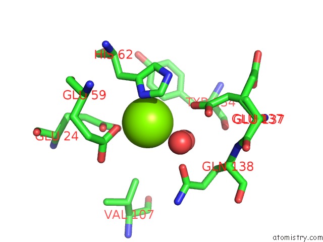
Mono view
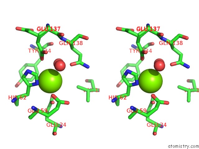
Stereo pair view
Mono view
Stereo pair view
|
|
A full contact list of Magnesium with other atoms in the Mg binding
site number 1 of Molecular Structure of Mouse Apoferritin Resolved at 2.7 Angstroms with the Glacios Cryo-Microscope within 5.0Å range:
|
Reference:
F.Hamdi,
C.Tuting,
D.A.Semchonok,
K.M.Visscher,
F.L.Kyrilis,
A.Meister,
I.Skalidis,
L.Schmidt,
C.Parthier,
M.T.Stubbs,
P.L.Kastritis.
2.7 Angstrom Cryo-Em Structure of Vitrified M. Musculus H-Chain Apoferritin From A Compact 200 Kev Cryo-Microscope. Plos One V. 15 32540 2020.
ISSN: ESSN 1932-6203
PubMed: 32374767
DOI: 10.1371/JOURNAL.PONE.0232540
Page generated: Tue Oct 1 17:56:32 2024
ISSN: ESSN 1932-6203
PubMed: 32374767
DOI: 10.1371/JOURNAL.PONE.0232540
Last articles
Fe in 2YXOFe in 2YRS
Fe in 2YXC
Fe in 2YNM
Fe in 2YVJ
Fe in 2YP1
Fe in 2YU2
Fe in 2YU1
Fe in 2YQB
Fe in 2YOO