Magnesium »
PDB 6sht-6stf »
6sqj »
Magnesium in PDB 6sqj: Crystal Structure of Glycoprotein D of Equine Herpesvirus Type 1
Protein crystallography data
The structure of Crystal Structure of Glycoprotein D of Equine Herpesvirus Type 1, PDB code: 6sqj
was solved by
V.Kremling,
B.Loll,
W.Azab,
N.Osterrieder,
I.Dahmani,
P.Chiantia,
M.Wahl,
with X-Ray Crystallography technique. A brief refinement statistics is given in the table below:
| Resolution Low / High (Å) | 49.80 / 2.25 |
| Space group | P 21 21 21 |
| Cell size a, b, c (Å), α, β, γ (°) | 71.860, 94.464, 101.296, 90.00, 90.00, 90.00 |
| R / Rfree (%) | 20.3 / 25.7 |
Magnesium Binding Sites:
The binding sites of Magnesium atom in the Crystal Structure of Glycoprotein D of Equine Herpesvirus Type 1
(pdb code 6sqj). This binding sites where shown within
5.0 Angstroms radius around Magnesium atom.
In total 2 binding sites of Magnesium where determined in the Crystal Structure of Glycoprotein D of Equine Herpesvirus Type 1, PDB code: 6sqj:
Jump to Magnesium binding site number: 1; 2;
In total 2 binding sites of Magnesium where determined in the Crystal Structure of Glycoprotein D of Equine Herpesvirus Type 1, PDB code: 6sqj:
Jump to Magnesium binding site number: 1; 2;
Magnesium binding site 1 out of 2 in 6sqj
Go back to
Magnesium binding site 1 out
of 2 in the Crystal Structure of Glycoprotein D of Equine Herpesvirus Type 1
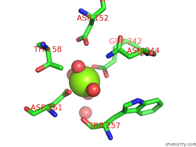
Mono view
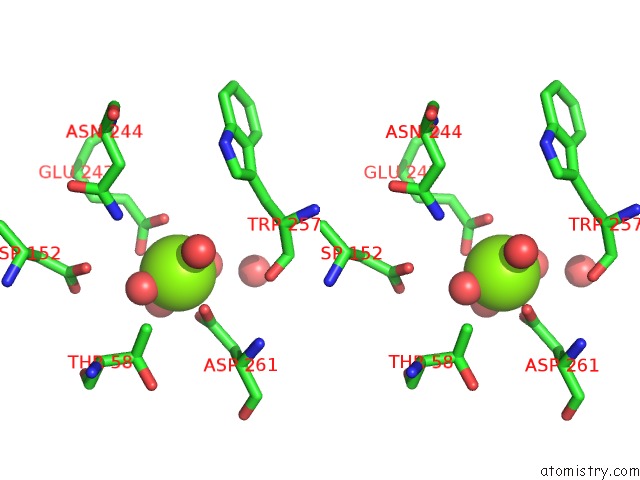
Stereo pair view
Mono view
Stereo pair view
A full contact list of Magnesium with other atoms in the Mg binding
site number 1 of Crystal Structure of Glycoprotein D of Equine Herpesvirus Type 1 within 5.0Å range:
|
Magnesium binding site 2 out of 2 in 6sqj
Go back to
Magnesium binding site 2 out
of 2 in the Crystal Structure of Glycoprotein D of Equine Herpesvirus Type 1
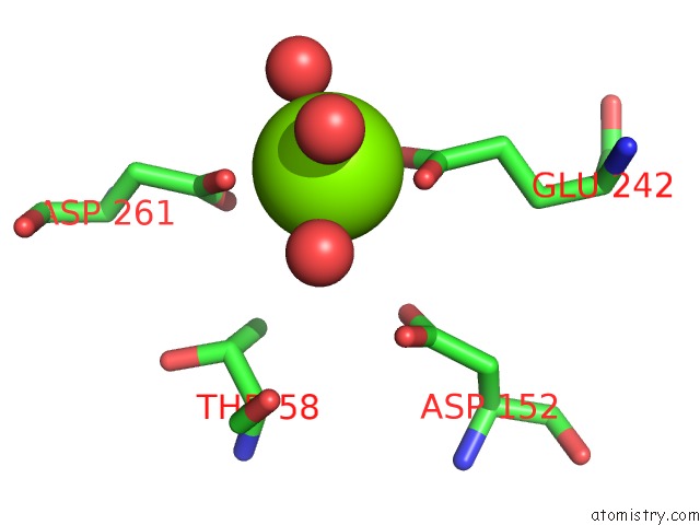
Mono view
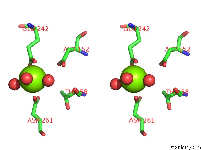
Stereo pair view
Mono view
Stereo pair view
A full contact list of Magnesium with other atoms in the Mg binding
site number 2 of Crystal Structure of Glycoprotein D of Equine Herpesvirus Type 1 within 5.0Å range:
|
Reference:
V.Kremling,
B.Loll,
W.Azab,
N.Osterrieder,
I.Dahmani,
P.Chiantia.
Crystal Structure of Glycoprotein D of Equine Herpesvirus Type 1 To Be Published.
Page generated: Tue Oct 1 18:02:08 2024
Last articles
Mg in 4JSTMg in 4JS0
Mg in 4JRY
Mg in 4JRC
Mg in 4JRP
Mg in 4JRN
Mg in 4JQP
Mg in 4JR7
Mg in 4JNX
Mg in 4JQL