Magnesium »
PDB 6stg-6t26 »
6sy1 »
Magnesium in PDB 6sy1: Crystal Structure of A Dehydrogenase
Enzymatic activity of Crystal Structure of A Dehydrogenase
All present enzymatic activity of Crystal Structure of A Dehydrogenase:
1.2.4.2;
1.2.4.2;
Protein crystallography data
The structure of Crystal Structure of A Dehydrogenase, PDB code: 6sy1
was solved by
G.A.Bezerra,
W.Foster,
L.Shrestha,
I.A.Pena,
J.Coker,
S.Kolker,
B.B.Nicola,
F.Von Delft,
A.Edwards,
C.Arrowsmith,
C.Bountra,
W.W.Yue,
Structuralgenomics Consortium (Sgc),
with X-Ray Crystallography technique. A brief refinement statistics is given in the table below:
| Resolution Low / High (Å) | 46.01 / 1.87 |
| Space group | P 1 |
| Cell size a, b, c (Å), α, β, γ (°) | 78.550, 81.220, 86.900, 63.43, 76.96, 72.06 |
| R / Rfree (%) | 19 / 23 |
Magnesium Binding Sites:
The binding sites of Magnesium atom in the Crystal Structure of A Dehydrogenase
(pdb code 6sy1). This binding sites where shown within
5.0 Angstroms radius around Magnesium atom.
In total 3 binding sites of Magnesium where determined in the Crystal Structure of A Dehydrogenase, PDB code: 6sy1:
Jump to Magnesium binding site number: 1; 2; 3;
In total 3 binding sites of Magnesium where determined in the Crystal Structure of A Dehydrogenase, PDB code: 6sy1:
Jump to Magnesium binding site number: 1; 2; 3;
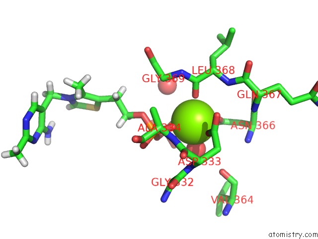
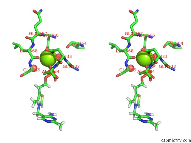
Magnesium binding site 1 out of 3 in 6sy1
Go back to
Magnesium binding site 1 out
of 3 in the Crystal Structure of A Dehydrogenase
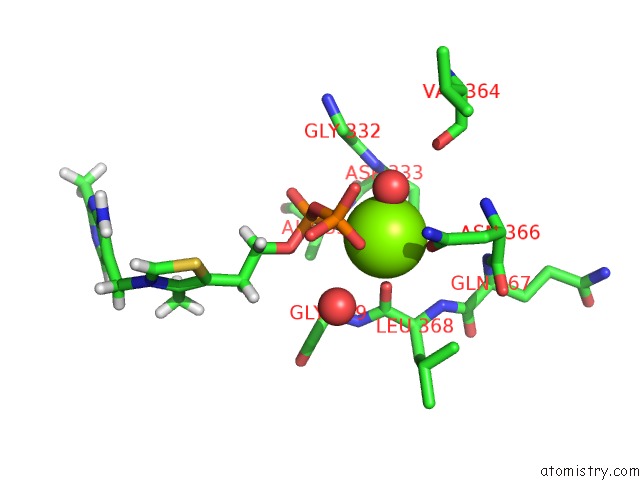
Mono view
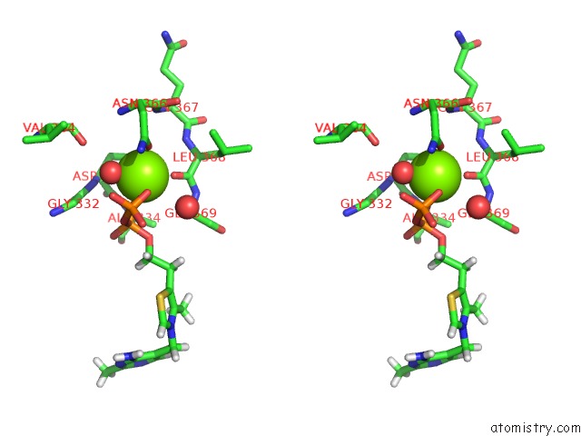
Stereo pair view
Mono view
Stereo pair view
A full contact list of Magnesium with other atoms in the Mg binding
site number 1 of Crystal Structure of A Dehydrogenase within 5.0Å range:
|
Magnesium binding site 2 out of 3 in 6sy1
Go back to
Magnesium binding site 2 out
of 3 in the Crystal Structure of A Dehydrogenase
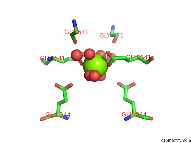
Mono view
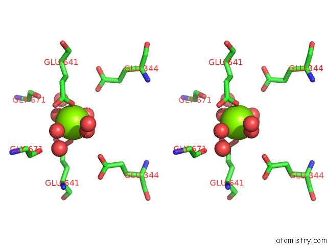
Stereo pair view
Mono view
Stereo pair view
A full contact list of Magnesium with other atoms in the Mg binding
site number 2 of Crystal Structure of A Dehydrogenase within 5.0Å range:
|
Magnesium binding site 3 out of 3 in 6sy1
Go back to
Magnesium binding site 3 out
of 3 in the Crystal Structure of A Dehydrogenase
Mono view
Stereo pair view
Mono view
Stereo pair view
A full contact list of Magnesium with other atoms in the Mg binding
site number 3 of Crystal Structure of A Dehydrogenase within 5.0Å range:
|
Reference:
G.A.Bezerra,
W.Foster,
L.Shrestha,
I.A.Pena,
J.Coker,
S.Kolker,
A.Edwards,
C.Arrowsmith,
C.Bountra,
W.Y.Wyatt.
Crystal Structure of Dehydrogenase E1 and Transketolase Domain-Containing Protein 1 (DHTKD1) To Be Published.
Page generated: Tue Oct 1 18:27:47 2024
Last articles
Mg in 4W5OMg in 4W5J
Mg in 4W5N
Mg in 4V2I
Mg in 4V3R
Mg in 4V26
Mg in 4V2G
Mg in 4V1T
Mg in 4V25
Mg in 4V1V