Magnesium »
PDB 6vzs-6wic »
6wfv »
Magnesium in PDB 6wfv: The Crystal Structure of A Collagen Galactosylhydroxylysyl Glucosyltransferase From Human
Enzymatic activity of The Crystal Structure of A Collagen Galactosylhydroxylysyl Glucosyltransferase From Human
All present enzymatic activity of The Crystal Structure of A Collagen Galactosylhydroxylysyl Glucosyltransferase From Human:
1.14.11.4; 2.4.1.50; 2.4.1.66;
1.14.11.4; 2.4.1.50; 2.4.1.66;
Protein crystallography data
The structure of The Crystal Structure of A Collagen Galactosylhydroxylysyl Glucosyltransferase From Human, PDB code: 6wfv
was solved by
H.-F.Guo,
C.-L.Tsai,
M.D.Miller,
G.N.Philips Jr.,
J.A.Tainer,
J.M.Kurie,
with X-Ray Crystallography technique. A brief refinement statistics is given in the table below:
| Resolution Low / High (Å) | 41.18 / 1.70 |
| Space group | P 32 2 1 |
| Cell size a, b, c (Å), α, β, γ (°) | 71.066, 71.066, 110.813, 90, 90, 120 |
| R / Rfree (%) | 15.2 / 17.2 |
Other elements in 6wfv:
The structure of The Crystal Structure of A Collagen Galactosylhydroxylysyl Glucosyltransferase From Human also contains other interesting chemical elements:
| Manganese | (Mn) | 1 atom |
Magnesium Binding Sites:
The binding sites of Magnesium atom in the The Crystal Structure of A Collagen Galactosylhydroxylysyl Glucosyltransferase From Human
(pdb code 6wfv). This binding sites where shown within
5.0 Angstroms radius around Magnesium atom.
In total 2 binding sites of Magnesium where determined in the The Crystal Structure of A Collagen Galactosylhydroxylysyl Glucosyltransferase From Human, PDB code: 6wfv:
Jump to Magnesium binding site number: 1; 2;
In total 2 binding sites of Magnesium where determined in the The Crystal Structure of A Collagen Galactosylhydroxylysyl Glucosyltransferase From Human, PDB code: 6wfv:
Jump to Magnesium binding site number: 1; 2;
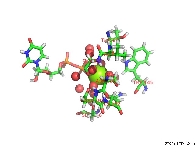
Magnesium binding site 1 out of 2 in 6wfv
Go back to
Magnesium binding site 1 out
of 2 in the The Crystal Structure of A Collagen Galactosylhydroxylysyl Glucosyltransferase From Human

Mono view

Stereo pair view
Mono view
Stereo pair view
A full contact list of Magnesium with other atoms in the Mg binding
site number 1 of The Crystal Structure of A Collagen Galactosylhydroxylysyl Glucosyltransferase From Human within 5.0Å range:
|
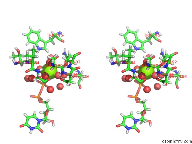
Magnesium binding site 2 out of 2 in 6wfv
Go back to
Magnesium binding site 2 out
of 2 in the The Crystal Structure of A Collagen Galactosylhydroxylysyl Glucosyltransferase From Human
Mono view
Stereo pair view
Mono view
Stereo pair view
A full contact list of Magnesium with other atoms in the Mg binding
site number 2 of The Crystal Structure of A Collagen Galactosylhydroxylysyl Glucosyltransferase From Human within 5.0Å range:
|
Reference:
H.-F.Guo,
C.-L.Tsai,
M.D.Miller,
G.N.Philips Jr.,
J.A.Tainer,
J.M.Kurie.
The Crystal Structure of A Collagen Galactosylhydroxylysyl Glucosyltransferase From Human To Be Published.
Page generated: Tue Oct 1 22:57:50 2024
Last articles
K in 6PQ7K in 6PNV
K in 6PIA
K in 6PID
K in 6PI1
K in 6PI8
K in 6PHT
K in 6PHR
K in 6PHZ
K in 6PCD