Magnesium »
PDB 6y0y-6yac »
6y44 »
Magnesium in PDB 6y44: 14-3-3 Sigma in Complex with Phosphorylated SOS1 Peptide
Protein crystallography data
The structure of 14-3-3 Sigma in Complex with Phosphorylated SOS1 Peptide, PDB code: 6y44
was solved by
A.Ballone,
R.A.Lau,
F.P.A.Zweipfenning,
C.Ottmann,
with X-Ray Crystallography technique. A brief refinement statistics is given in the table below:
| Resolution Low / High (Å) | 29.81 / 1.71 |
| Space group | C 2 2 21 |
| Cell size a, b, c (Å), α, β, γ (°) | 81.769, 111.627, 62.792, 90.00, 90.00, 90.00 |
| R / Rfree (%) | 17.4 / 20.2 |
Other elements in 6y44:
The structure of 14-3-3 Sigma in Complex with Phosphorylated SOS1 Peptide also contains other interesting chemical elements:
| Calcium | (Ca) | 1 atom |
| Chlorine | (Cl) | 4 atoms |
Magnesium Binding Sites:
The binding sites of Magnesium atom in the 14-3-3 Sigma in Complex with Phosphorylated SOS1 Peptide
(pdb code 6y44). This binding sites where shown within
5.0 Angstroms radius around Magnesium atom.
In total 3 binding sites of Magnesium where determined in the 14-3-3 Sigma in Complex with Phosphorylated SOS1 Peptide, PDB code: 6y44:
Jump to Magnesium binding site number: 1; 2; 3;
In total 3 binding sites of Magnesium where determined in the 14-3-3 Sigma in Complex with Phosphorylated SOS1 Peptide, PDB code: 6y44:
Jump to Magnesium binding site number: 1; 2; 3;
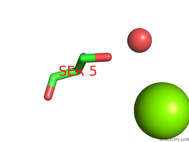
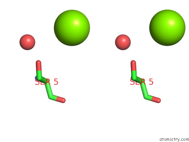
Magnesium binding site 1 out of 3 in 6y44
Go back to
Magnesium binding site 1 out
of 3 in the 14-3-3 Sigma in Complex with Phosphorylated SOS1 Peptide
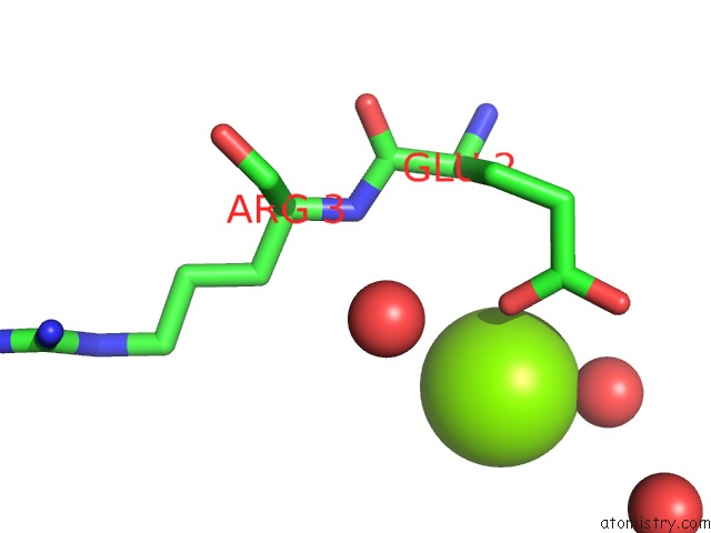
Mono view
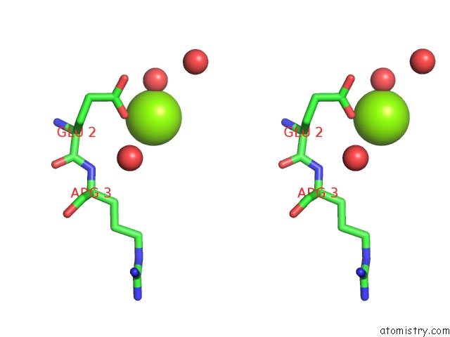
Stereo pair view
Mono view
Stereo pair view
A full contact list of Magnesium with other atoms in the Mg binding
site number 1 of 14-3-3 Sigma in Complex with Phosphorylated SOS1 Peptide within 5.0Å range:
|
Magnesium binding site 2 out of 3 in 6y44
Go back to
Magnesium binding site 2 out
of 3 in the 14-3-3 Sigma in Complex with Phosphorylated SOS1 Peptide
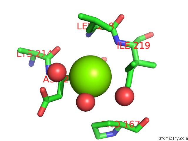
Mono view
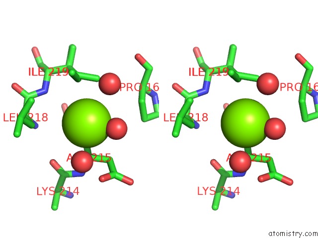
Stereo pair view
Mono view
Stereo pair view
A full contact list of Magnesium with other atoms in the Mg binding
site number 2 of 14-3-3 Sigma in Complex with Phosphorylated SOS1 Peptide within 5.0Å range:
|
Magnesium binding site 3 out of 3 in 6y44
Go back to
Magnesium binding site 3 out
of 3 in the 14-3-3 Sigma in Complex with Phosphorylated SOS1 Peptide
Mono view
Stereo pair view
Mono view
Stereo pair view
A full contact list of Magnesium with other atoms in the Mg binding
site number 3 of 14-3-3 Sigma in Complex with Phosphorylated SOS1 Peptide within 5.0Å range:
|
Reference:
A.Ballone,
R.A.Lau,
F.P.A.Zweipfenning,
C.Ottmann.
A New Soaking Procedure For X-Ray Crystallographic Structural Determination of Protein-Peptide Complexes. Acta Crystallogr.,Sect.F V. 76 501 2020.
ISSN: ESSN 2053-230X
PubMed: 33006579
DOI: 10.1107/S2053230X2001122X
Page generated: Wed Oct 2 00:08:55 2024
ISSN: ESSN 2053-230X
PubMed: 33006579
DOI: 10.1107/S2053230X2001122X
Last articles
Mg in 3TVKMg in 3TVB
Mg in 3TVA
Mg in 3TUY
Mg in 3TTE
Mg in 3TTF
Mg in 3TTD
Mg in 3TTC
Mg in 3TSU
Mg in 3TSO