Magnesium »
PDB 6zup-7a1x »
7a0c »
Magnesium in PDB 7a0c: X-Ray Structure of Nika From Escherichia Coli in Complex with Fe-6- ME2-Bpmcn
Protein crystallography data
The structure of X-Ray Structure of Nika From Escherichia Coli in Complex with Fe-6- ME2-Bpmcn, PDB code: 7a0c
was solved by
C.Cavazza,
S.Menage,
with X-Ray Crystallography technique. A brief refinement statistics is given in the table below:
| Resolution Low / High (Å) | 43.88 / 1.90 |
| Space group | P 21 21 21 |
| Cell size a, b, c (Å), α, β, γ (°) | 86.809, 93.796, 124.222, 90.00, 90.00, 90.00 |
| R / Rfree (%) | 18.2 / 22.2 |
Other elements in 7a0c:
The structure of X-Ray Structure of Nika From Escherichia Coli in Complex with Fe-6- ME2-Bpmcn also contains other interesting chemical elements:
| Iron | (Fe) | 2 atoms |
| Chlorine | (Cl) | 1 atom |
Magnesium Binding Sites:
The binding sites of Magnesium atom in the X-Ray Structure of Nika From Escherichia Coli in Complex with Fe-6- ME2-Bpmcn
(pdb code 7a0c). This binding sites where shown within
5.0 Angstroms radius around Magnesium atom.
In total 5 binding sites of Magnesium where determined in the X-Ray Structure of Nika From Escherichia Coli in Complex with Fe-6- ME2-Bpmcn, PDB code: 7a0c:
Jump to Magnesium binding site number: 1; 2; 3; 4; 5;
In total 5 binding sites of Magnesium where determined in the X-Ray Structure of Nika From Escherichia Coli in Complex with Fe-6- ME2-Bpmcn, PDB code: 7a0c:
Jump to Magnesium binding site number: 1; 2; 3; 4; 5;
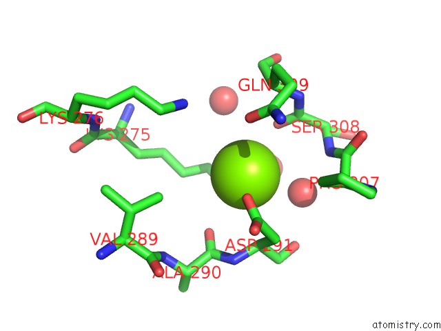
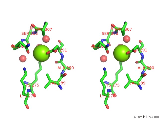
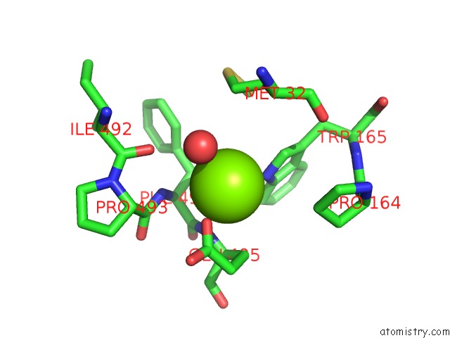
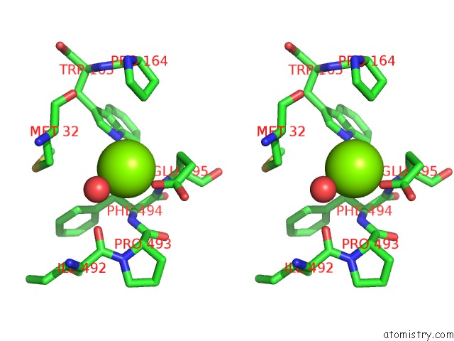
Magnesium binding site 1 out of 5 in 7a0c
Go back to
Magnesium binding site 1 out
of 5 in the X-Ray Structure of Nika From Escherichia Coli in Complex with Fe-6- ME2-Bpmcn
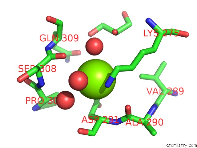
Mono view
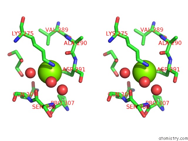
Stereo pair view
Mono view
Stereo pair view
A full contact list of Magnesium with other atoms in the Mg binding
site number 1 of X-Ray Structure of Nika From Escherichia Coli in Complex with Fe-6- ME2-Bpmcn within 5.0Å range:
|
Magnesium binding site 2 out of 5 in 7a0c
Go back to
Magnesium binding site 2 out
of 5 in the X-Ray Structure of Nika From Escherichia Coli in Complex with Fe-6- ME2-Bpmcn
Mono view
Stereo pair view
Mono view
Stereo pair view
A full contact list of Magnesium with other atoms in the Mg binding
site number 2 of X-Ray Structure of Nika From Escherichia Coli in Complex with Fe-6- ME2-Bpmcn within 5.0Å range:
|
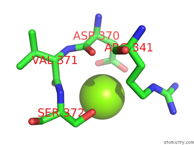
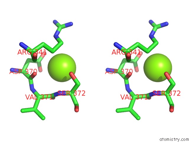
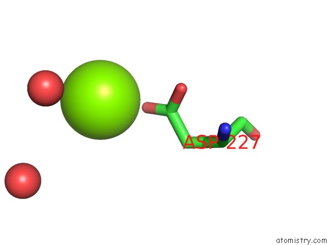
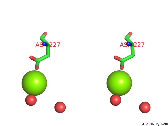
Magnesium binding site 3 out of 5 in 7a0c
Go back to
Magnesium binding site 3 out
of 5 in the X-Ray Structure of Nika From Escherichia Coli in Complex with Fe-6- ME2-Bpmcn
Mono view
Stereo pair view
Mono view
Stereo pair view
A full contact list of Magnesium with other atoms in the Mg binding
site number 3 of X-Ray Structure of Nika From Escherichia Coli in Complex with Fe-6- ME2-Bpmcn within 5.0Å range:
|
Magnesium binding site 4 out of 5 in 7a0c
Go back to
Magnesium binding site 4 out
of 5 in the X-Ray Structure of Nika From Escherichia Coli in Complex with Fe-6- ME2-Bpmcn
Mono view
Stereo pair view
Mono view
Stereo pair view
A full contact list of Magnesium with other atoms in the Mg binding
site number 4 of X-Ray Structure of Nika From Escherichia Coli in Complex with Fe-6- ME2-Bpmcn within 5.0Å range:
|
Magnesium binding site 5 out of 5 in 7a0c
Go back to
Magnesium binding site 5 out
of 5 in the X-Ray Structure of Nika From Escherichia Coli in Complex with Fe-6- ME2-Bpmcn
Mono view
Stereo pair view
Mono view
Stereo pair view
A full contact list of Magnesium with other atoms in the Mg binding
site number 5 of X-Ray Structure of Nika From Escherichia Coli in Complex with Fe-6- ME2-Bpmcn within 5.0Å range:
|
Reference:
S.Lopez,
C.Marchi-Delapierre,
C.Cavazza,
S.Menage.
A Selective Sulfide Oxidation Catalyzed By Heterogeneous Artificial Metalloenzymes Iron@Nika. Chemistry 2020.
ISSN: ISSN 0947-6539
PubMed: 33079395
DOI: 10.1002/CHEM.202003746
Page generated: Wed Oct 2 03:22:05 2024
ISSN: ISSN 0947-6539
PubMed: 33079395
DOI: 10.1002/CHEM.202003746
Last articles
Fe in 8FB0Fe in 8F6N
Fe in 8F5W
Fe in 8F61
Fe in 8F9N
Fe in 8F9J
Fe in 8F9I
Fe in 8F9H
Fe in 8F91
Fe in 8F6T