Magnesium »
PDB 6zup-7a1x »
7a17 »
Magnesium in PDB 7a17: Crystal Structure of the 5-Phosphatase Domain of SYNAPTOJANIN1 Bound to Its Substrate DIC8-Pi(3,4,5)P3 in Complex with A Nanobody
Enzymatic activity of Crystal Structure of the 5-Phosphatase Domain of SYNAPTOJANIN1 Bound to Its Substrate DIC8-Pi(3,4,5)P3 in Complex with A Nanobody
All present enzymatic activity of Crystal Structure of the 5-Phosphatase Domain of SYNAPTOJANIN1 Bound to Its Substrate DIC8-Pi(3,4,5)P3 in Complex with A Nanobody:
3.1.3.36;
3.1.3.36;
Protein crystallography data
The structure of Crystal Structure of the 5-Phosphatase Domain of SYNAPTOJANIN1 Bound to Its Substrate DIC8-Pi(3,4,5)P3 in Complex with A Nanobody, PDB code: 7a17
was solved by
J.Paesmans,
C.Galicia,
E.Martin,
W.Versees,
with X-Ray Crystallography technique. A brief refinement statistics is given in the table below:
| Resolution Low / High (Å) | 87.39 / 2.73 |
| Space group | C 1 2 1 |
| Cell size a, b, c (Å), α, β, γ (°) | 169.323, 109.205, 100.902, 90, 120.62, 90 |
| R / Rfree (%) | 19.6 / 25.7 |
Magnesium Binding Sites:
The binding sites of Magnesium atom in the Crystal Structure of the 5-Phosphatase Domain of SYNAPTOJANIN1 Bound to Its Substrate DIC8-Pi(3,4,5)P3 in Complex with A Nanobody
(pdb code 7a17). This binding sites where shown within
5.0 Angstroms radius around Magnesium atom.
In total 2 binding sites of Magnesium where determined in the Crystal Structure of the 5-Phosphatase Domain of SYNAPTOJANIN1 Bound to Its Substrate DIC8-Pi(3,4,5)P3 in Complex with A Nanobody, PDB code: 7a17:
Jump to Magnesium binding site number: 1; 2;
In total 2 binding sites of Magnesium where determined in the Crystal Structure of the 5-Phosphatase Domain of SYNAPTOJANIN1 Bound to Its Substrate DIC8-Pi(3,4,5)P3 in Complex with A Nanobody, PDB code: 7a17:
Jump to Magnesium binding site number: 1; 2;
Magnesium binding site 1 out of 2 in 7a17
Go back to
Magnesium binding site 1 out
of 2 in the Crystal Structure of the 5-Phosphatase Domain of SYNAPTOJANIN1 Bound to Its Substrate DIC8-Pi(3,4,5)P3 in Complex with A Nanobody

Mono view
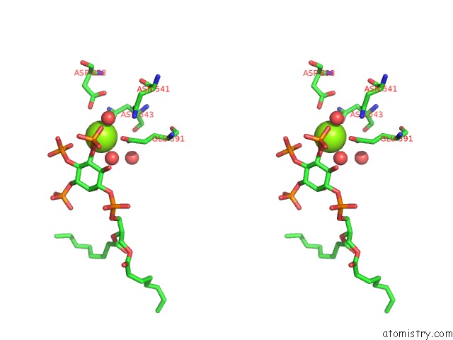
Stereo pair view
Mono view
Stereo pair view
A full contact list of Magnesium with other atoms in the Mg binding
site number 1 of Crystal Structure of the 5-Phosphatase Domain of SYNAPTOJANIN1 Bound to Its Substrate DIC8-Pi(3,4,5)P3 in Complex with A Nanobody within 5.0Å range:
|
Magnesium binding site 2 out of 2 in 7a17
Go back to
Magnesium binding site 2 out
of 2 in the Crystal Structure of the 5-Phosphatase Domain of SYNAPTOJANIN1 Bound to Its Substrate DIC8-Pi(3,4,5)P3 in Complex with A Nanobody

Mono view

Stereo pair view
Mono view
Stereo pair view
A full contact list of Magnesium with other atoms in the Mg binding
site number 2 of Crystal Structure of the 5-Phosphatase Domain of SYNAPTOJANIN1 Bound to Its Substrate DIC8-Pi(3,4,5)P3 in Complex with A Nanobody within 5.0Å range:
|
Reference:
J.Paesmans,
E.Martin,
B.Deckers,
M.Berghmans,
R.Sethi,
Y.Loeys,
E.Pardon,
J.Steyaert,
P.Verstreken,
C.Galicia,
W.Versees.
A Structure of Substrate-Bound SYNAPTOJANIN1 Provides New Insights in Its Mechanism and the Effect of Disease Mutations. Elife V. 9 2020.
ISSN: ESSN 2050-084X
PubMed: 33349335
DOI: 10.7554/ELIFE.64922
Page generated: Wed Oct 2 04:21:07 2024
ISSN: ESSN 2050-084X
PubMed: 33349335
DOI: 10.7554/ELIFE.64922
Last articles
K in 6YD5K in 6YD1
K in 6Y72
K in 6YAA
K in 6Y3A
K in 6XYB
K in 6XRQ
K in 6XV4
K in 6XUP
K in 6XU0