Magnesium »
PDB 7ae1-7ap2 »
7aie »
Magnesium in PDB 7aie: Crystal Structure of A Truncated Form of the KLC1-Tpr Domain ([A1-B5] Fragment) - Monoclinic Crystal Form
Protein crystallography data
The structure of Crystal Structure of A Truncated Form of the KLC1-Tpr Domain ([A1-B5] Fragment) - Monoclinic Crystal Form, PDB code: 7aie
was solved by
J.Menetrey,
P.Llinas,
with X-Ray Crystallography technique. A brief refinement statistics is given in the table below:
| Resolution Low / High (Å) | 46.33 / 3.29 |
| Space group | P 1 21 1 |
| Cell size a, b, c (Å), α, β, γ (°) | 71.49, 72.71, 110.87, 90, 90.05, 90 |
| R / Rfree (%) | 20.1 / 25 |
Magnesium Binding Sites:
The binding sites of Magnesium atom in the Crystal Structure of A Truncated Form of the KLC1-Tpr Domain ([A1-B5] Fragment) - Monoclinic Crystal Form
(pdb code 7aie). This binding sites where shown within
5.0 Angstroms radius around Magnesium atom.
In total 2 binding sites of Magnesium where determined in the Crystal Structure of A Truncated Form of the KLC1-Tpr Domain ([A1-B5] Fragment) - Monoclinic Crystal Form, PDB code: 7aie:
Jump to Magnesium binding site number: 1; 2;
In total 2 binding sites of Magnesium where determined in the Crystal Structure of A Truncated Form of the KLC1-Tpr Domain ([A1-B5] Fragment) - Monoclinic Crystal Form, PDB code: 7aie:
Jump to Magnesium binding site number: 1; 2;
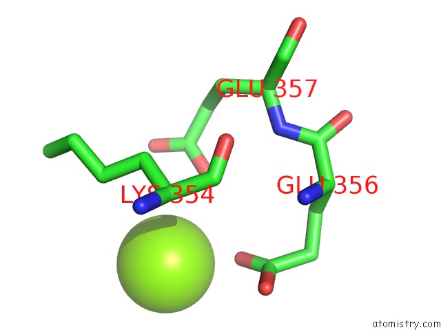
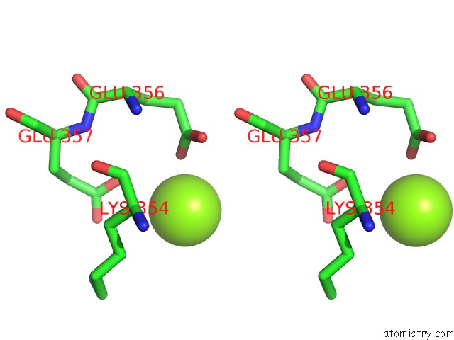
Magnesium binding site 1 out of 2 in 7aie
Go back to
Magnesium binding site 1 out
of 2 in the Crystal Structure of A Truncated Form of the KLC1-Tpr Domain ([A1-B5] Fragment) - Monoclinic Crystal Form

Mono view

Stereo pair view
Mono view
Stereo pair view
A full contact list of Magnesium with other atoms in the Mg binding
site number 1 of Crystal Structure of A Truncated Form of the KLC1-Tpr Domain ([A1-B5] Fragment) - Monoclinic Crystal Form within 5.0Å range:
|
Magnesium binding site 2 out of 2 in 7aie
Go back to
Magnesium binding site 2 out
of 2 in the Crystal Structure of A Truncated Form of the KLC1-Tpr Domain ([A1-B5] Fragment) - Monoclinic Crystal Form
Mono view
Stereo pair view
Mono view
Stereo pair view
A full contact list of Magnesium with other atoms in the Mg binding
site number 2 of Crystal Structure of A Truncated Form of the KLC1-Tpr Domain ([A1-B5] Fragment) - Monoclinic Crystal Form within 5.0Å range:
|
Reference:
J.Menetrey,
P.Llinas.
Structural Investigations of the Dynamics of the Tpr Domain of Kinesin Light Chain To Be Published.
Page generated: Thu Aug 14 01:13:20 2025
Last articles
Mg in 7BOEMg in 7BGI
Mg in 7BLX
Mg in 7BLZ
Mg in 7BOD
Mg in 7BNR
Mg in 7BNK
Mg in 7BMC
Mg in 7BM9
Mg in 7BM8