Magnesium »
PDB 7b2c-7bdk »
7b9h »
Magnesium in PDB 7b9h: Crystal Structure of the PDE4D Catalytic Domain in Complex with Gebr- 42A
Enzymatic activity of Crystal Structure of the PDE4D Catalytic Domain in Complex with Gebr- 42A
All present enzymatic activity of Crystal Structure of the PDE4D Catalytic Domain in Complex with Gebr- 42A:
3.1.4.53;
3.1.4.53;
Protein crystallography data
The structure of Crystal Structure of the PDE4D Catalytic Domain in Complex with Gebr- 42A, PDB code: 7b9h
was solved by
A.Torretta,
S.Abbate,
E.Parisini,
with X-Ray Crystallography technique. A brief refinement statistics is given in the table below:
| Resolution Low / High (Å) | 54.13 / 1.50 |
| Space group | P 21 21 21 |
| Cell size a, b, c (Å), α, β, γ (°) | 64.748, 98.628, 119.821, 90, 90, 90 |
| R / Rfree (%) | 19.4 / 22.1 |
Other elements in 7b9h:
The structure of Crystal Structure of the PDE4D Catalytic Domain in Complex with Gebr- 42A also contains other interesting chemical elements:
| Zinc | (Zn) | 2 atoms |
Magnesium Binding Sites:
The binding sites of Magnesium atom in the Crystal Structure of the PDE4D Catalytic Domain in Complex with Gebr- 42A
(pdb code 7b9h). This binding sites where shown within
5.0 Angstroms radius around Magnesium atom.
In total 8 binding sites of Magnesium where determined in the Crystal Structure of the PDE4D Catalytic Domain in Complex with Gebr- 42A, PDB code: 7b9h:
Jump to Magnesium binding site number: 1; 2; 3; 4; 5; 6; 7; 8;
In total 8 binding sites of Magnesium where determined in the Crystal Structure of the PDE4D Catalytic Domain in Complex with Gebr- 42A, PDB code: 7b9h:
Jump to Magnesium binding site number: 1; 2; 3; 4; 5; 6; 7; 8;
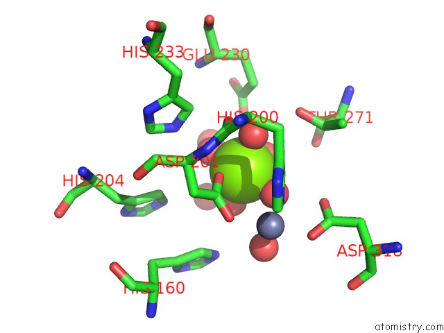
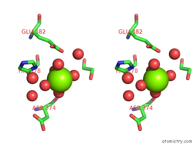
Magnesium binding site 1 out of 8 in 7b9h
Go back to
Magnesium binding site 1 out
of 8 in the Crystal Structure of the PDE4D Catalytic Domain in Complex with Gebr- 42A

Mono view
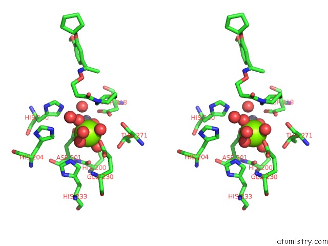
Stereo pair view
Mono view
Stereo pair view
A full contact list of Magnesium with other atoms in the Mg binding
site number 1 of Crystal Structure of the PDE4D Catalytic Domain in Complex with Gebr- 42A within 5.0Å range:
|
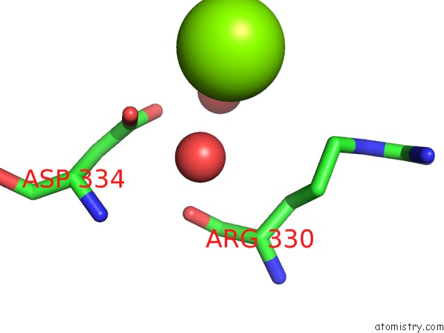
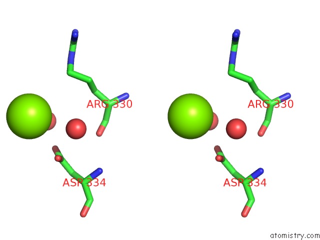
Magnesium binding site 2 out of 8 in 7b9h
Go back to
Magnesium binding site 2 out
of 8 in the Crystal Structure of the PDE4D Catalytic Domain in Complex with Gebr- 42A
Mono view

Stereo pair view
Mono view
Stereo pair view
A full contact list of Magnesium with other atoms in the Mg binding
site number 2 of Crystal Structure of the PDE4D Catalytic Domain in Complex with Gebr- 42A within 5.0Å range:
|
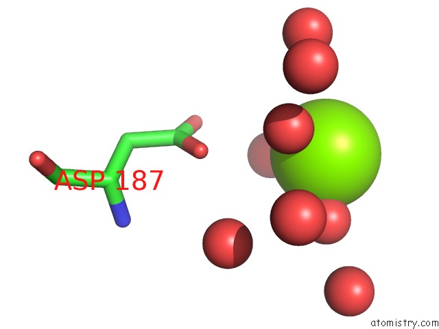
Magnesium binding site 3 out of 8 in 7b9h
Go back to
Magnesium binding site 3 out
of 8 in the Crystal Structure of the PDE4D Catalytic Domain in Complex with Gebr- 42A

Mono view
Stereo pair view
Mono view
Stereo pair view
A full contact list of Magnesium with other atoms in the Mg binding
site number 3 of Crystal Structure of the PDE4D Catalytic Domain in Complex with Gebr- 42A within 5.0Å range:
|
Magnesium binding site 4 out of 8 in 7b9h
Go back to
Magnesium binding site 4 out
of 8 in the Crystal Structure of the PDE4D Catalytic Domain in Complex with Gebr- 42A

Mono view
Stereo pair view
Mono view
Stereo pair view
A full contact list of Magnesium with other atoms in the Mg binding
site number 4 of Crystal Structure of the PDE4D Catalytic Domain in Complex with Gebr- 42A within 5.0Å range:
|
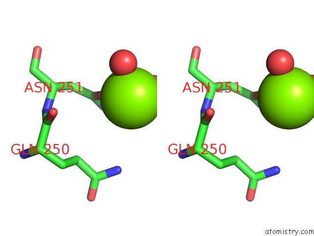
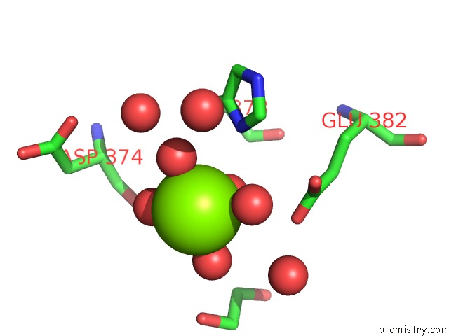
Magnesium binding site 5 out of 8 in 7b9h
Go back to
Magnesium binding site 5 out
of 8 in the Crystal Structure of the PDE4D Catalytic Domain in Complex with Gebr- 42A

Mono view

Stereo pair view
Mono view
Stereo pair view
A full contact list of Magnesium with other atoms in the Mg binding
site number 5 of Crystal Structure of the PDE4D Catalytic Domain in Complex with Gebr- 42A within 5.0Å range:
|
Magnesium binding site 6 out of 8 in 7b9h
Go back to
Magnesium binding site 6 out
of 8 in the Crystal Structure of the PDE4D Catalytic Domain in Complex with Gebr- 42A
Mono view

Stereo pair view
Mono view
Stereo pair view
A full contact list of Magnesium with other atoms in the Mg binding
site number 6 of Crystal Structure of the PDE4D Catalytic Domain in Complex with Gebr- 42A within 5.0Å range:
|
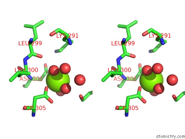
Magnesium binding site 7 out of 8 in 7b9h
Go back to
Magnesium binding site 7 out
of 8 in the Crystal Structure of the PDE4D Catalytic Domain in Complex with Gebr- 42A
Mono view
Stereo pair view
Mono view
Stereo pair view
A full contact list of Magnesium with other atoms in the Mg binding
site number 7 of Crystal Structure of the PDE4D Catalytic Domain in Complex with Gebr- 42A within 5.0Å range:
|
Magnesium binding site 8 out of 8 in 7b9h
Go back to
Magnesium binding site 8 out
of 8 in the Crystal Structure of the PDE4D Catalytic Domain in Complex with Gebr- 42A
Mono view
Stereo pair view
Mono view
Stereo pair view
A full contact list of Magnesium with other atoms in the Mg binding
site number 8 of Crystal Structure of the PDE4D Catalytic Domain in Complex with Gebr- 42A within 5.0Å range:
|
Reference:
C.Brullo,
F.Rapetti,
S.Abbate,
T.Prosdocimi,
A.Torretta,
M.Semrau,
M.Massa,
S.Alfei,
P.Storici,
E.Parisini,
O.Bruno.
Design, Synthesis, Biological Evaluation and Structural Characterization of Novel Gebr Library PDE4D Inhibitors Eur.J.Med.Chem. 2021.
ISSN: ISSN 0223-5234
DOI: 10.1016/J.EJMECH.2021.113638
Page generated: Thu Aug 14 01:24:38 2025
ISSN: ISSN 0223-5234
DOI: 10.1016/J.EJMECH.2021.113638
Last articles
Zn in 7VUWZn in 7VVR
Zn in 7VWJ
Zn in 7VTX
Zn in 7VTV
Zn in 7VTW
Zn in 7VD8
Zn in 7VSP
Zn in 7VSE
Zn in 7VSL