Magnesium »
PDB 7c7j-7cji »
7cj5 »
Magnesium in PDB 7cj5: Crystal Structure of Homo Dimeric D-Allulose 3-Epimerase From Methylomonas Sp. in Complex with D-Fructose
Protein crystallography data
The structure of Crystal Structure of Homo Dimeric D-Allulose 3-Epimerase From Methylomonas Sp. in Complex with D-Fructose, PDB code: 7cj5
was solved by
H.Yoshida,
A.Yoshihara,
S.Kamitori,
with X-Ray Crystallography technique. A brief refinement statistics is given in the table below:
| Resolution Low / High (Å) | 19.94 / 1.80 |
| Space group | P 21 21 21 |
| Cell size a, b, c (Å), α, β, γ (°) | 45.42, 70.54, 140.22, 90, 90, 90 |
| R / Rfree (%) | 12.6 / 15.9 |
Other elements in 7cj5:
The structure of Crystal Structure of Homo Dimeric D-Allulose 3-Epimerase From Methylomonas Sp. in Complex with D-Fructose also contains other interesting chemical elements:
| Manganese | (Mn) | 2 atoms |
Magnesium Binding Sites:
The binding sites of Magnesium atom in the Crystal Structure of Homo Dimeric D-Allulose 3-Epimerase From Methylomonas Sp. in Complex with D-Fructose
(pdb code 7cj5). This binding sites where shown within
5.0 Angstroms radius around Magnesium atom.
In total 2 binding sites of Magnesium where determined in the Crystal Structure of Homo Dimeric D-Allulose 3-Epimerase From Methylomonas Sp. in Complex with D-Fructose, PDB code: 7cj5:
Jump to Magnesium binding site number: 1; 2;
In total 2 binding sites of Magnesium where determined in the Crystal Structure of Homo Dimeric D-Allulose 3-Epimerase From Methylomonas Sp. in Complex with D-Fructose, PDB code: 7cj5:
Jump to Magnesium binding site number: 1; 2;
Magnesium binding site 1 out of 2 in 7cj5
Go back to
Magnesium binding site 1 out
of 2 in the Crystal Structure of Homo Dimeric D-Allulose 3-Epimerase From Methylomonas Sp. in Complex with D-Fructose
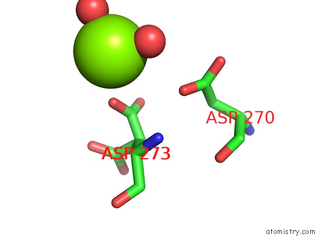
Mono view
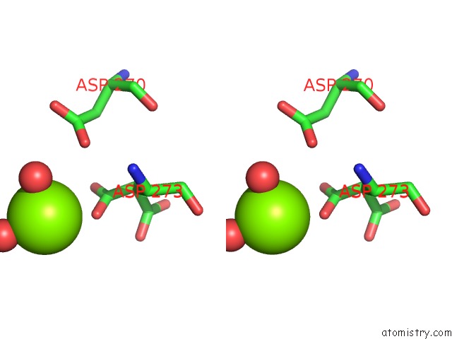
Stereo pair view
Mono view
Stereo pair view
A full contact list of Magnesium with other atoms in the Mg binding
site number 1 of Crystal Structure of Homo Dimeric D-Allulose 3-Epimerase From Methylomonas Sp. in Complex with D-Fructose within 5.0Å range:
|
Magnesium binding site 2 out of 2 in 7cj5
Go back to
Magnesium binding site 2 out
of 2 in the Crystal Structure of Homo Dimeric D-Allulose 3-Epimerase From Methylomonas Sp. in Complex with D-Fructose
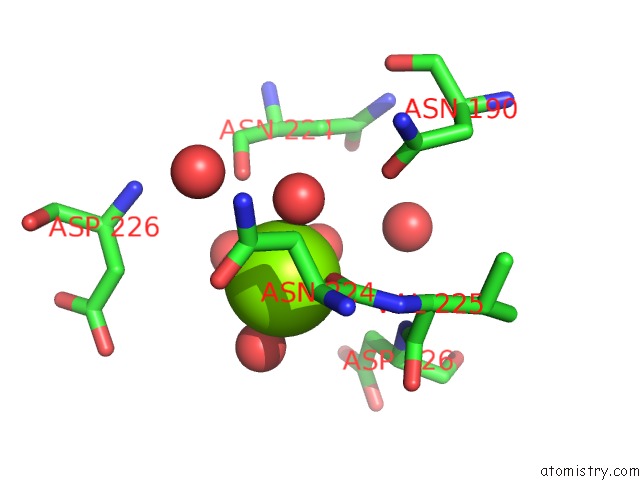
Mono view
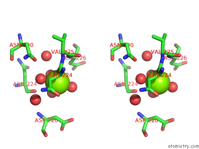
Stereo pair view
Mono view
Stereo pair view
A full contact list of Magnesium with other atoms in the Mg binding
site number 2 of Crystal Structure of Homo Dimeric D-Allulose 3-Epimerase From Methylomonas Sp. in Complex with D-Fructose within 5.0Å range:
|
Reference:
H.Yoshida,
A.Yoshihara,
S.Kato,
S.Mochizuki,
K.Akimitsu,
K.Izumori,
S.Kamitori.
Crystal Structure of A Novel Homodimeric L-Ribulose 3-Epimerase From Methylomonus Sp. Febs Open Bio 2021.
ISSN: ESSN 2211-5463
PubMed: 33838083
DOI: 10.1002/2211-5463.13159
Page generated: Wed Oct 2 14:04:32 2024
ISSN: ESSN 2211-5463
PubMed: 33838083
DOI: 10.1002/2211-5463.13159
Last articles
K in 4IZNK in 4JAH
K in 4J1R
K in 4J6D
K in 4J6C
K in 4J6B
K in 4J2T
K in 4IX2
K in 4IRU
K in 4IQ0