Magnesium »
PDB 7cu9-7d8g »
7d7y »
Magnesium in PDB 7d7y: Crystal Structure of the DOMAIN1 of Nad+ Riboswitch with Adenosine Triphosphate (Atp)
Protein crystallography data
The structure of Crystal Structure of the DOMAIN1 of Nad+ Riboswitch with Adenosine Triphosphate (Atp), PDB code: 7d7y
was solved by
H.Chen,
A.M.Ren,
with X-Ray Crystallography technique. A brief refinement statistics is given in the table below:
| Resolution Low / High (Å) | 28.26 / 2.80 |
| Space group | I 2 2 2 |
| Cell size a, b, c (Å), α, β, γ (°) | 56.528, 57.703, 196.758, 90.00, 90.00, 90.00 |
| R / Rfree (%) | 22 / 24 |
Magnesium Binding Sites:
The binding sites of Magnesium atom in the Crystal Structure of the DOMAIN1 of Nad+ Riboswitch with Adenosine Triphosphate (Atp)
(pdb code 7d7y). This binding sites where shown within
5.0 Angstroms radius around Magnesium atom.
In total 8 binding sites of Magnesium where determined in the Crystal Structure of the DOMAIN1 of Nad+ Riboswitch with Adenosine Triphosphate (Atp), PDB code: 7d7y:
Jump to Magnesium binding site number: 1; 2; 3; 4; 5; 6; 7; 8;
In total 8 binding sites of Magnesium where determined in the Crystal Structure of the DOMAIN1 of Nad+ Riboswitch with Adenosine Triphosphate (Atp), PDB code: 7d7y:
Jump to Magnesium binding site number: 1; 2; 3; 4; 5; 6; 7; 8;
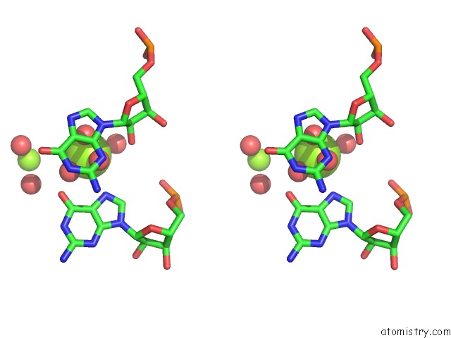
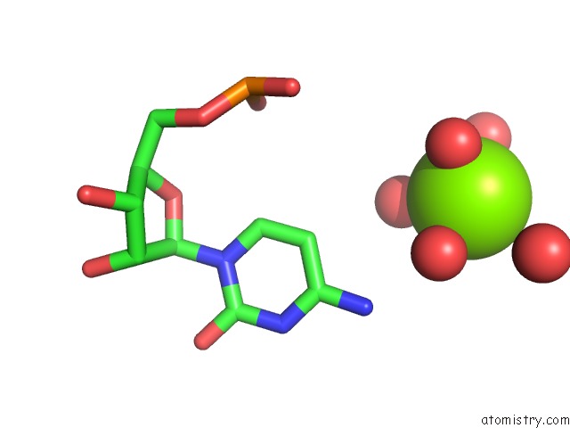
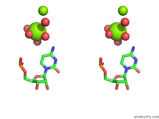
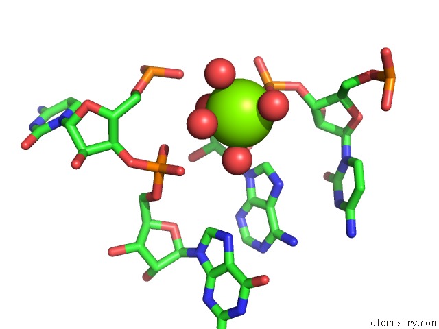
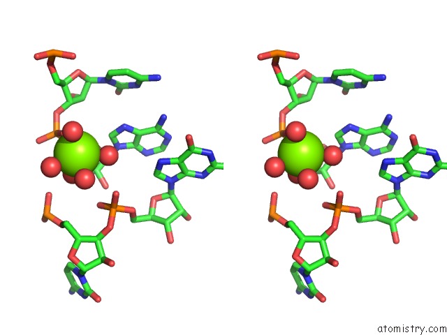
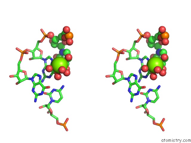
Magnesium binding site 1 out of 8 in 7d7y
Go back to
Magnesium binding site 1 out
of 8 in the Crystal Structure of the DOMAIN1 of Nad+ Riboswitch with Adenosine Triphosphate (Atp)
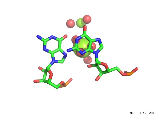
Mono view
Stereo pair view
Mono view
Stereo pair view
A full contact list of Magnesium with other atoms in the Mg binding
site number 1 of Crystal Structure of the DOMAIN1 of Nad+ Riboswitch with Adenosine Triphosphate (Atp) within 5.0Å range:
|
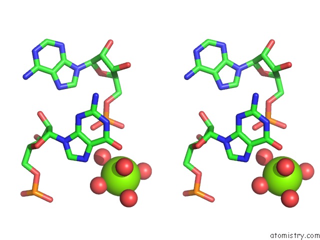
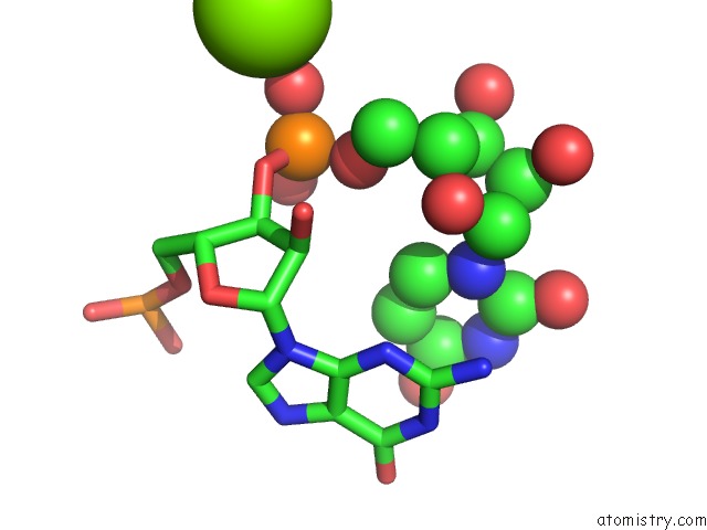
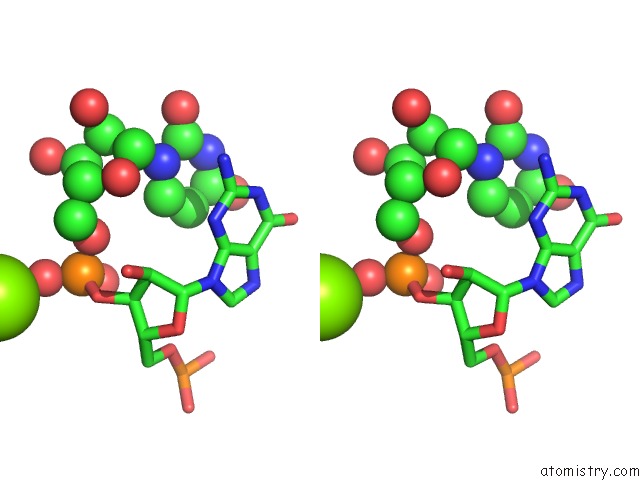
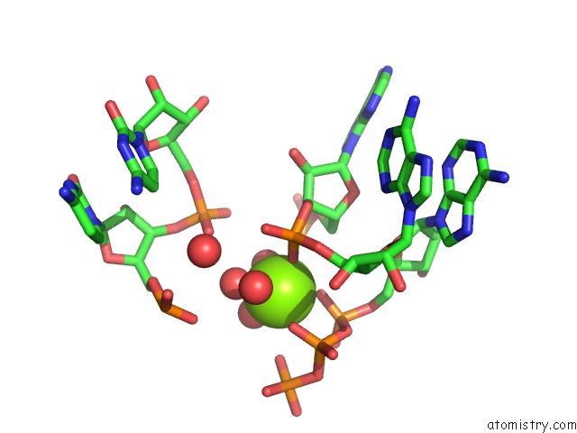
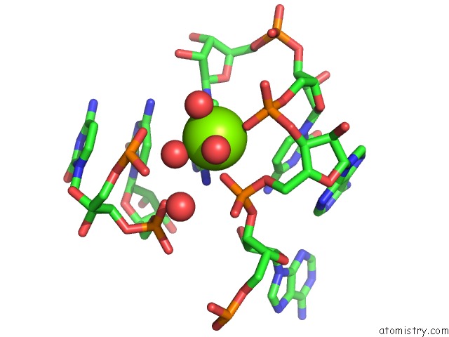
Magnesium binding site 2 out of 8 in 7d7y
Go back to
Magnesium binding site 2 out
of 8 in the Crystal Structure of the DOMAIN1 of Nad+ Riboswitch with Adenosine Triphosphate (Atp)
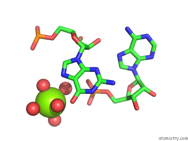
Mono view
Stereo pair view
Mono view
Stereo pair view
A full contact list of Magnesium with other atoms in the Mg binding
site number 2 of Crystal Structure of the DOMAIN1 of Nad+ Riboswitch with Adenosine Triphosphate (Atp) within 5.0Å range:
|
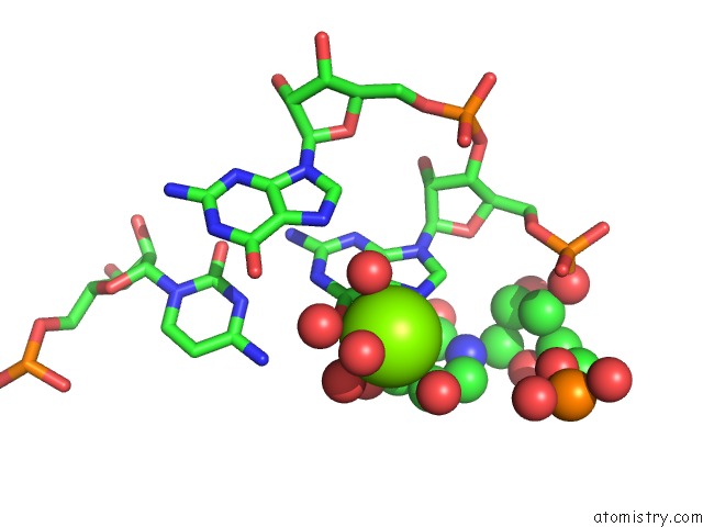
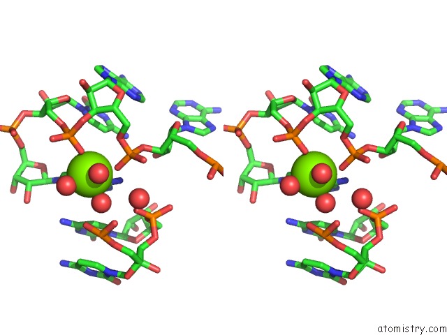
Magnesium binding site 3 out of 8 in 7d7y
Go back to
Magnesium binding site 3 out
of 8 in the Crystal Structure of the DOMAIN1 of Nad+ Riboswitch with Adenosine Triphosphate (Atp)
Mono view
Stereo pair view
Mono view
Stereo pair view
A full contact list of Magnesium with other atoms in the Mg binding
site number 3 of Crystal Structure of the DOMAIN1 of Nad+ Riboswitch with Adenosine Triphosphate (Atp) within 5.0Å range:
|
Magnesium binding site 4 out of 8 in 7d7y
Go back to
Magnesium binding site 4 out
of 8 in the Crystal Structure of the DOMAIN1 of Nad+ Riboswitch with Adenosine Triphosphate (Atp)
Mono view
Stereo pair view
Mono view
Stereo pair view
A full contact list of Magnesium with other atoms in the Mg binding
site number 4 of Crystal Structure of the DOMAIN1 of Nad+ Riboswitch with Adenosine Triphosphate (Atp) within 5.0Å range:
|
Magnesium binding site 5 out of 8 in 7d7y
Go back to
Magnesium binding site 5 out
of 8 in the Crystal Structure of the DOMAIN1 of Nad+ Riboswitch with Adenosine Triphosphate (Atp)
Mono view
Stereo pair view
Mono view
Stereo pair view
A full contact list of Magnesium with other atoms in the Mg binding
site number 5 of Crystal Structure of the DOMAIN1 of Nad+ Riboswitch with Adenosine Triphosphate (Atp) within 5.0Å range:
|
Magnesium binding site 6 out of 8 in 7d7y
Go back to
Magnesium binding site 6 out
of 8 in the Crystal Structure of the DOMAIN1 of Nad+ Riboswitch with Adenosine Triphosphate (Atp)
Mono view

Stereo pair view
Mono view
Stereo pair view
A full contact list of Magnesium with other atoms in the Mg binding
site number 6 of Crystal Structure of the DOMAIN1 of Nad+ Riboswitch with Adenosine Triphosphate (Atp) within 5.0Å range:
|
Magnesium binding site 7 out of 8 in 7d7y
Go back to
Magnesium binding site 7 out
of 8 in the Crystal Structure of the DOMAIN1 of Nad+ Riboswitch with Adenosine Triphosphate (Atp)
Mono view
Stereo pair view
Mono view
Stereo pair view
A full contact list of Magnesium with other atoms in the Mg binding
site number 7 of Crystal Structure of the DOMAIN1 of Nad+ Riboswitch with Adenosine Triphosphate (Atp) within 5.0Å range:
|
Magnesium binding site 8 out of 8 in 7d7y
Go back to
Magnesium binding site 8 out
of 8 in the Crystal Structure of the DOMAIN1 of Nad+ Riboswitch with Adenosine Triphosphate (Atp)
Mono view
Stereo pair view
Mono view
Stereo pair view
A full contact list of Magnesium with other atoms in the Mg binding
site number 8 of Crystal Structure of the DOMAIN1 of Nad+ Riboswitch with Adenosine Triphosphate (Atp) within 5.0Å range:
|
Reference:
H.Chen,
M.Egger,
X.Xu,
L.Flemmich,
O.Krasheninina,
A.Sun,
R.Micura,
A.Ren.
Structural Distinctions Between Nad+ Riboswitch Domains 1 and 2 Determine Differential Folding and Ligand Binding. Nucleic Acids Res. 2020.
ISSN: ESSN 1362-4962
PubMed: 33170270
DOI: 10.1093/NAR/GKAA1029
Page generated: Wed Oct 2 14:46:08 2024
ISSN: ESSN 1362-4962
PubMed: 33170270
DOI: 10.1093/NAR/GKAA1029
Last articles
Mg in 5KFUMg in 5KFT
Mg in 5KFP
Mg in 5KFN
Mg in 5KF6
Mg in 5KF7
Mg in 5KF0
Mg in 5KEU
Mg in 5KDL
Mg in 5KAP