Magnesium »
PDB 7ent-7evi »
7eul »
Magnesium in PDB 7eul: Crystal Structure of C86H-H196S Mutant of N(Omega)-Hydroxy-L-Arginine Hydrolase
Enzymatic activity of Crystal Structure of C86H-H196S Mutant of N(Omega)-Hydroxy-L-Arginine Hydrolase
All present enzymatic activity of Crystal Structure of C86H-H196S Mutant of N(Omega)-Hydroxy-L-Arginine Hydrolase:
3.5.3.25;
3.5.3.25;
Protein crystallography data
The structure of Crystal Structure of C86H-H196S Mutant of N(Omega)-Hydroxy-L-Arginine Hydrolase, PDB code: 7eul
was solved by
K.Oda,
Y.Matoba,
with X-Ray Crystallography technique. A brief refinement statistics is given in the table below:
| Resolution Low / High (Å) | 41.93 / 1.45 |
| Space group | P 1 21 1 |
| Cell size a, b, c (Å), α, β, γ (°) | 42.499, 50.343, 52.941, 90, 99.42, 90 |
| R / Rfree (%) | 16.9 / 19.3 |
Other elements in 7eul:
The structure of Crystal Structure of C86H-H196S Mutant of N(Omega)-Hydroxy-L-Arginine Hydrolase also contains other interesting chemical elements:
| Manganese | (Mn) | 3 atoms |
Magnesium Binding Sites:
The binding sites of Magnesium atom in the Crystal Structure of C86H-H196S Mutant of N(Omega)-Hydroxy-L-Arginine Hydrolase
(pdb code 7eul). This binding sites where shown within
5.0 Angstroms radius around Magnesium atom.
In total 2 binding sites of Magnesium where determined in the Crystal Structure of C86H-H196S Mutant of N(Omega)-Hydroxy-L-Arginine Hydrolase, PDB code: 7eul:
Jump to Magnesium binding site number: 1; 2;
In total 2 binding sites of Magnesium where determined in the Crystal Structure of C86H-H196S Mutant of N(Omega)-Hydroxy-L-Arginine Hydrolase, PDB code: 7eul:
Jump to Magnesium binding site number: 1; 2;
Magnesium binding site 1 out of 2 in 7eul
Go back to
Magnesium binding site 1 out
of 2 in the Crystal Structure of C86H-H196S Mutant of N(Omega)-Hydroxy-L-Arginine Hydrolase
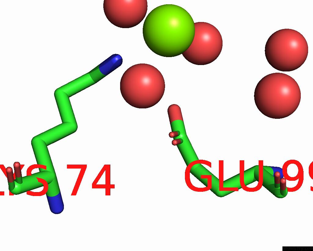
Mono view
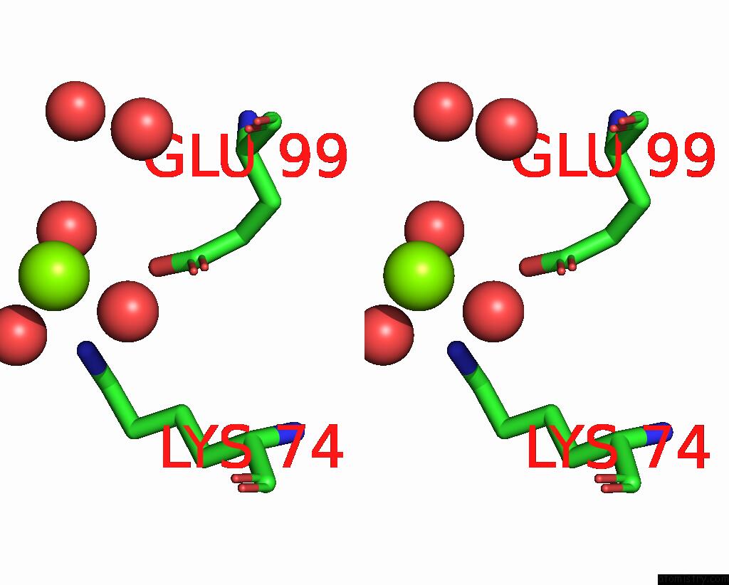
Stereo pair view
Mono view
Stereo pair view
A full contact list of Magnesium with other atoms in the Mg binding
site number 1 of Crystal Structure of C86H-H196S Mutant of N(Omega)-Hydroxy-L-Arginine Hydrolase within 5.0Å range:
|
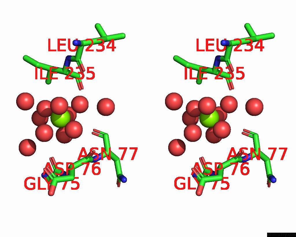
Magnesium binding site 2 out of 2 in 7eul
Go back to
Magnesium binding site 2 out
of 2 in the Crystal Structure of C86H-H196S Mutant of N(Omega)-Hydroxy-L-Arginine Hydrolase
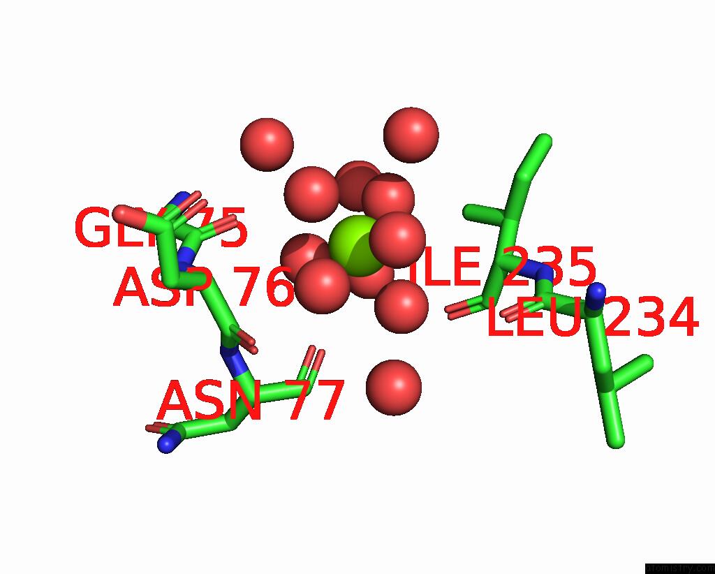
Mono view
Stereo pair view
Mono view
Stereo pair view
A full contact list of Magnesium with other atoms in the Mg binding
site number 2 of Crystal Structure of C86H-H196S Mutant of N(Omega)-Hydroxy-L-Arginine Hydrolase within 5.0Å range:
|
Reference:
K.Oda,
T.Sakaguchi,
Y.Matoba.
Catalytic Mechanism of Dcsb: Arginase Framework Used For Hydrolyzing Its Inhibitor. Protein Sci. V. 31 E4338 2022.
ISSN: ESSN 1469-896X
PubMed: 35634777
DOI: 10.1002/PRO.4338
Page generated: Wed Oct 2 20:59:08 2024
ISSN: ESSN 1469-896X
PubMed: 35634777
DOI: 10.1002/PRO.4338
Last articles
K in 4RBQK in 4R8I
K in 4R4V
K in 4R7J
K in 4R1L
K in 4R47
K in 4R45
K in 4R33
K in 4R2C
K in 4QXG