Magnesium »
PDB 7ftr-7jig »
7jhi »
Magnesium in PDB 7jhi: Structure of Human Beta 1,3-N-Acetylglucosaminyltransferase 2 Iodide- Derivative
Enzymatic activity of Structure of Human Beta 1,3-N-Acetylglucosaminyltransferase 2 Iodide- Derivative
All present enzymatic activity of Structure of Human Beta 1,3-N-Acetylglucosaminyltransferase 2 Iodide- Derivative:
2.4.1.149;
2.4.1.149;
Protein crystallography data
The structure of Structure of Human Beta 1,3-N-Acetylglucosaminyltransferase 2 Iodide- Derivative, PDB code: 7jhi
was solved by
Y.Hao,
X.Huang,
with X-Ray Crystallography technique. A brief refinement statistics is given in the table below:
| Resolution Low / High (Å) | 47.33 / 2.50 |
| Space group | P 1 21 1 |
| Cell size a, b, c (Å), α, β, γ (°) | 67.431, 81.385, 157.138, 90.00, 98.63, 90.00 |
| R / Rfree (%) | 20.2 / 24.8 |
Other elements in 7jhi:
The structure of Structure of Human Beta 1,3-N-Acetylglucosaminyltransferase 2 Iodide- Derivative also contains other interesting chemical elements:
| Iodine | (I) | 41 atoms |
| Chlorine | (Cl) | 5 atoms |
Magnesium Binding Sites:
The binding sites of Magnesium atom in the Structure of Human Beta 1,3-N-Acetylglucosaminyltransferase 2 Iodide- Derivative
(pdb code 7jhi). This binding sites where shown within
5.0 Angstroms radius around Magnesium atom.
In total 3 binding sites of Magnesium where determined in the Structure of Human Beta 1,3-N-Acetylglucosaminyltransferase 2 Iodide- Derivative, PDB code: 7jhi:
Jump to Magnesium binding site number: 1; 2; 3;
In total 3 binding sites of Magnesium where determined in the Structure of Human Beta 1,3-N-Acetylglucosaminyltransferase 2 Iodide- Derivative, PDB code: 7jhi:
Jump to Magnesium binding site number: 1; 2; 3;
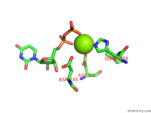
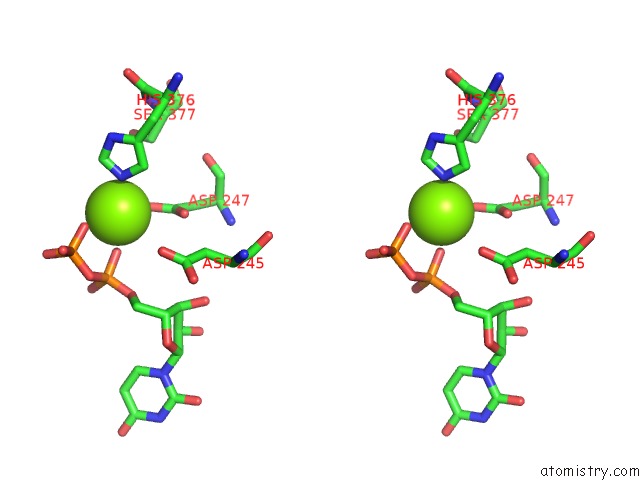
Magnesium binding site 1 out of 3 in 7jhi
Go back to
Magnesium binding site 1 out
of 3 in the Structure of Human Beta 1,3-N-Acetylglucosaminyltransferase 2 Iodide- Derivative
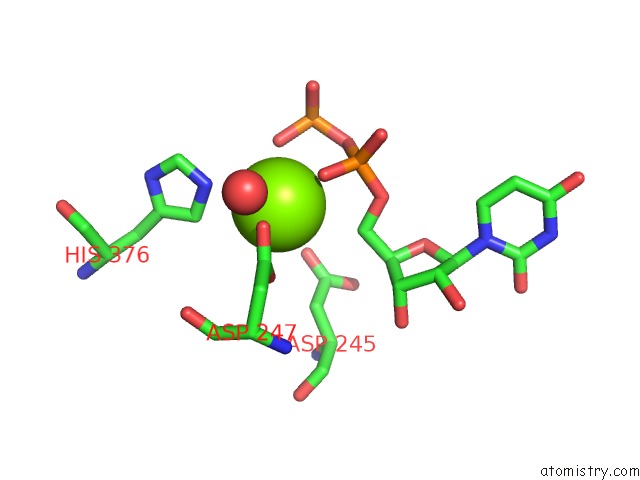
Mono view
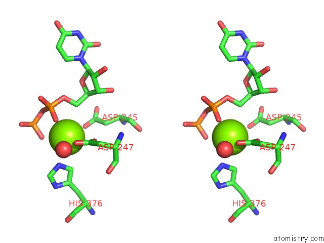
Stereo pair view
Mono view
Stereo pair view
A full contact list of Magnesium with other atoms in the Mg binding
site number 1 of Structure of Human Beta 1,3-N-Acetylglucosaminyltransferase 2 Iodide- Derivative within 5.0Å range:
|
Magnesium binding site 2 out of 3 in 7jhi
Go back to
Magnesium binding site 2 out
of 3 in the Structure of Human Beta 1,3-N-Acetylglucosaminyltransferase 2 Iodide- Derivative
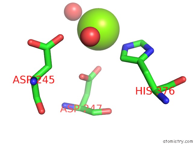
Mono view
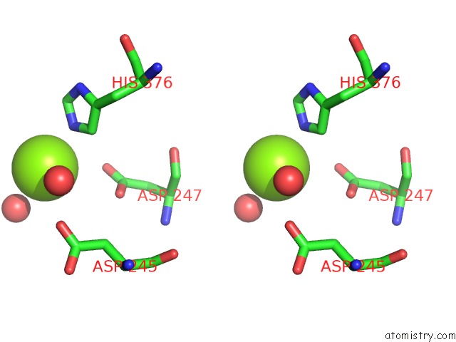
Stereo pair view
Mono view
Stereo pair view
A full contact list of Magnesium with other atoms in the Mg binding
site number 2 of Structure of Human Beta 1,3-N-Acetylglucosaminyltransferase 2 Iodide- Derivative within 5.0Å range:
|
Magnesium binding site 3 out of 3 in 7jhi
Go back to
Magnesium binding site 3 out
of 3 in the Structure of Human Beta 1,3-N-Acetylglucosaminyltransferase 2 Iodide- Derivative
Mono view
Stereo pair view
Mono view
Stereo pair view
A full contact list of Magnesium with other atoms in the Mg binding
site number 3 of Structure of Human Beta 1,3-N-Acetylglucosaminyltransferase 2 Iodide- Derivative within 5.0Å range:
|
Reference:
Y.Hao,
A.Crequer-Grandhomme,
N.Javier,
A.Singh,
H.Chen,
P.Manzanillo,
M.C.Lo,
X.Huang.
Structures and Mechanism of Human Glycosyltransferase Beta 1,3-N-Acetylglucosaminyltransferase 2 (B3GNT2), An Important Player in Immune Homeostasis. J.Biol.Chem. 2020.
ISSN: ESSN 1083-351X
PubMed: 33158990
DOI: 10.1074/JBC.RA120.015306
Page generated: Thu Aug 14 07:51:40 2025
ISSN: ESSN 1083-351X
PubMed: 33158990
DOI: 10.1074/JBC.RA120.015306
Last articles
Mn in 9LJUMn in 9LJW
Mn in 9LJS
Mn in 9LJR
Mn in 9LJT
Mn in 9LJV
Mg in 9UA2
Mg in 9R96
Mg in 9VM1
Mg in 9P01