Magnesium »
PDB 7m99-7miu »
7mbh »
Magnesium in PDB 7mbh: Structure of Human Enolase 2 in Complex with Phosphoserine
Enzymatic activity of Structure of Human Enolase 2 in Complex with Phosphoserine
All present enzymatic activity of Structure of Human Enolase 2 in Complex with Phosphoserine:
4.2.1.11;
4.2.1.11;
Protein crystallography data
The structure of Structure of Human Enolase 2 in Complex with Phosphoserine, PDB code: 7mbh
was solved by
P.G.Leonard,
K.G.Hicks,
J.Rutter,
with X-Ray Crystallography technique. A brief refinement statistics is given in the table below:
| Resolution Low / High (Å) | 42.39 / 2.10 |
| Space group | P 21 21 21 |
| Cell size a, b, c (Å), α, β, γ (°) | 68.05, 108.38, 117.929, 90, 90, 90 |
| R / Rfree (%) | 18 / 23.5 |
Magnesium Binding Sites:
The binding sites of Magnesium atom in the Structure of Human Enolase 2 in Complex with Phosphoserine
(pdb code 7mbh). This binding sites where shown within
5.0 Angstroms radius around Magnesium atom.
In total 2 binding sites of Magnesium where determined in the Structure of Human Enolase 2 in Complex with Phosphoserine, PDB code: 7mbh:
Jump to Magnesium binding site number: 1; 2;
In total 2 binding sites of Magnesium where determined in the Structure of Human Enolase 2 in Complex with Phosphoserine, PDB code: 7mbh:
Jump to Magnesium binding site number: 1; 2;
Magnesium binding site 1 out of 2 in 7mbh
Go back to
Magnesium binding site 1 out
of 2 in the Structure of Human Enolase 2 in Complex with Phosphoserine
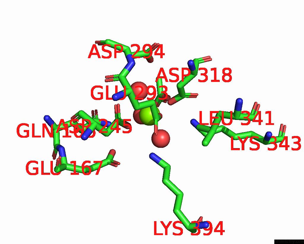
Mono view
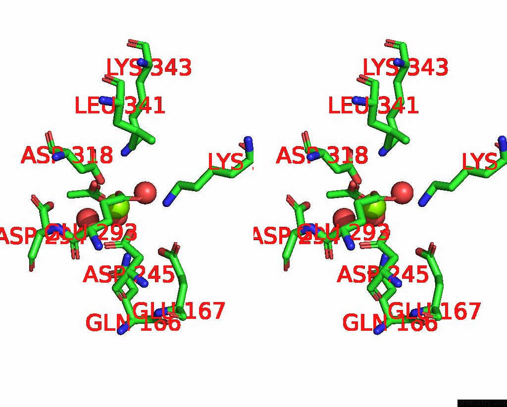
Stereo pair view
Mono view
Stereo pair view
A full contact list of Magnesium with other atoms in the Mg binding
site number 1 of Structure of Human Enolase 2 in Complex with Phosphoserine within 5.0Å range:
|
Magnesium binding site 2 out of 2 in 7mbh
Go back to
Magnesium binding site 2 out
of 2 in the Structure of Human Enolase 2 in Complex with Phosphoserine
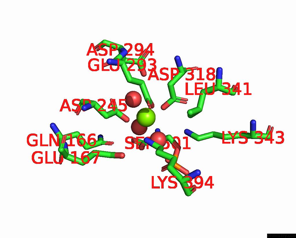
Mono view
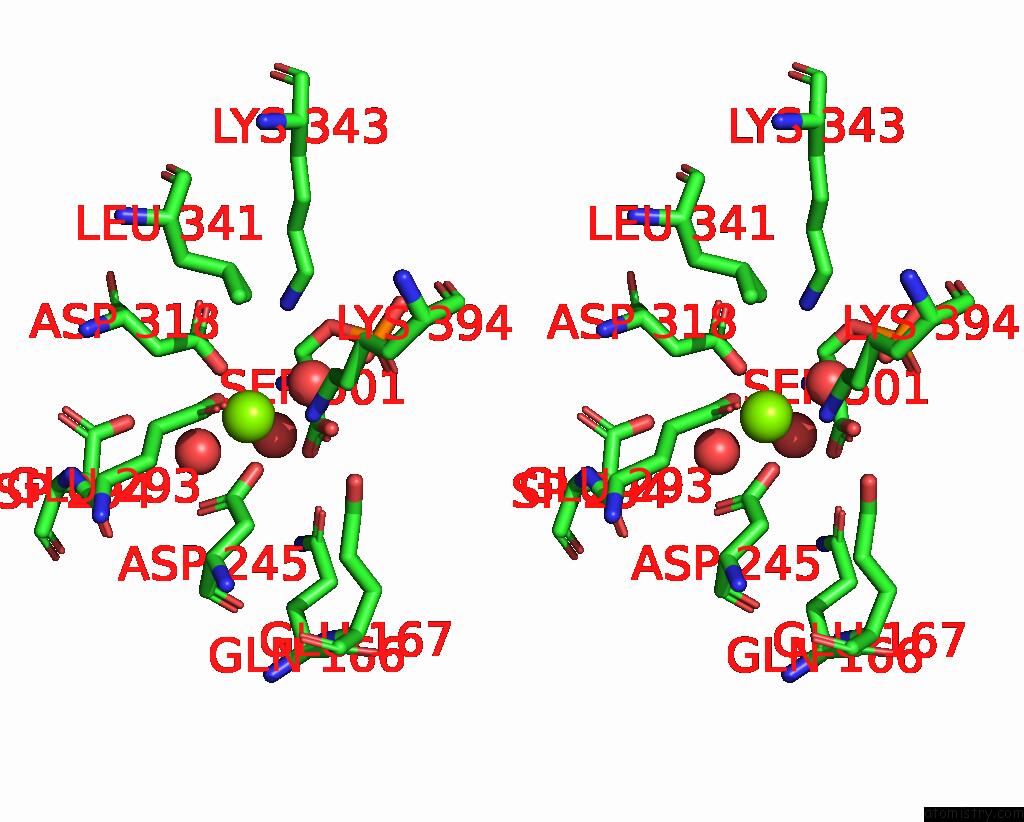
Stereo pair view
Mono view
Stereo pair view
A full contact list of Magnesium with other atoms in the Mg binding
site number 2 of Structure of Human Enolase 2 in Complex with Phosphoserine within 5.0Å range:
|
Reference:
K.G.Hick,
P.G.Leonard,
J.Rutter.
The Protein-Metabolite Interactome of Central Carbon Metabolism Reveals Novel Regulation of Pyruvate Metabolism To Be Published.
Page generated: Thu Aug 14 10:12:21 2025
Last articles
Mg in 7YQ7Mg in 7YQ2
Mg in 7YXG
Mg in 7YWM
Mg in 7YWA
Mg in 7YV1
Mg in 7YUZ
Mg in 7YUA
Mg in 7YSX
Mg in 7YSE