Magnesium »
PDB 7m99-7miu »
7mgn »
Magnesium in PDB 7mgn: Crystal Structure of F501H/H506E Variant of 2-Ketopropyl Coenzyme M Oxidoreductase/Carboxylase (2-Kpcc) From Xanthobacter Autotrophicus
Enzymatic activity of Crystal Structure of F501H/H506E Variant of 2-Ketopropyl Coenzyme M Oxidoreductase/Carboxylase (2-Kpcc) From Xanthobacter Autotrophicus
All present enzymatic activity of Crystal Structure of F501H/H506E Variant of 2-Ketopropyl Coenzyme M Oxidoreductase/Carboxylase (2-Kpcc) From Xanthobacter Autotrophicus:
1.8.1.5;
1.8.1.5;
Protein crystallography data
The structure of Crystal Structure of F501H/H506E Variant of 2-Ketopropyl Coenzyme M Oxidoreductase/Carboxylase (2-Kpcc) From Xanthobacter Autotrophicus, PDB code: 7mgn
was solved by
O.A.Zadvornyy,
G.Prussia,
J.W.Peters,
with X-Ray Crystallography technique. A brief refinement statistics is given in the table below:
| Resolution Low / High (Å) | 37.66 / 1.80 |
| Space group | P 1 21 1 |
| Cell size a, b, c (Å), α, β, γ (°) | 87.006, 60.111, 105.14, 90, 100.52, 90 |
| R / Rfree (%) | 14.1 / 18.9 |
Magnesium Binding Sites:
The binding sites of Magnesium atom in the Crystal Structure of F501H/H506E Variant of 2-Ketopropyl Coenzyme M Oxidoreductase/Carboxylase (2-Kpcc) From Xanthobacter Autotrophicus
(pdb code 7mgn). This binding sites where shown within
5.0 Angstroms radius around Magnesium atom.
In total 2 binding sites of Magnesium where determined in the Crystal Structure of F501H/H506E Variant of 2-Ketopropyl Coenzyme M Oxidoreductase/Carboxylase (2-Kpcc) From Xanthobacter Autotrophicus, PDB code: 7mgn:
Jump to Magnesium binding site number: 1; 2;
In total 2 binding sites of Magnesium where determined in the Crystal Structure of F501H/H506E Variant of 2-Ketopropyl Coenzyme M Oxidoreductase/Carboxylase (2-Kpcc) From Xanthobacter Autotrophicus, PDB code: 7mgn:
Jump to Magnesium binding site number: 1; 2;
Magnesium binding site 1 out of 2 in 7mgn
Go back to
Magnesium binding site 1 out
of 2 in the Crystal Structure of F501H/H506E Variant of 2-Ketopropyl Coenzyme M Oxidoreductase/Carboxylase (2-Kpcc) From Xanthobacter Autotrophicus
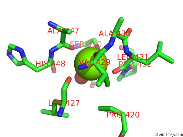
Mono view
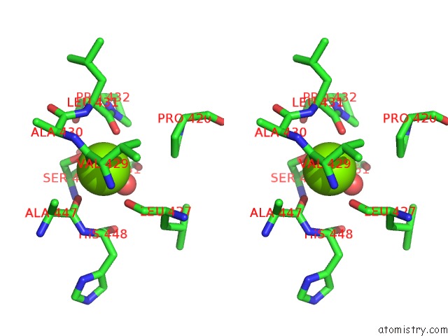
Stereo pair view
Mono view
Stereo pair view
A full contact list of Magnesium with other atoms in the Mg binding
site number 1 of Crystal Structure of F501H/H506E Variant of 2-Ketopropyl Coenzyme M Oxidoreductase/Carboxylase (2-Kpcc) From Xanthobacter Autotrophicus within 5.0Å range:
|
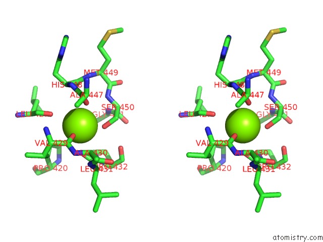
Magnesium binding site 2 out of 2 in 7mgn
Go back to
Magnesium binding site 2 out
of 2 in the Crystal Structure of F501H/H506E Variant of 2-Ketopropyl Coenzyme M Oxidoreductase/Carboxylase (2-Kpcc) From Xanthobacter Autotrophicus
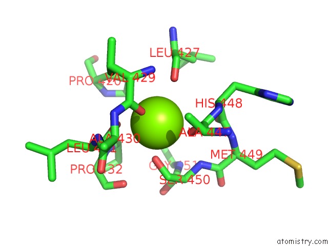
Mono view
Stereo pair view
Mono view
Stereo pair view
A full contact list of Magnesium with other atoms in the Mg binding
site number 2 of Crystal Structure of F501H/H506E Variant of 2-Ketopropyl Coenzyme M Oxidoreductase/Carboxylase (2-Kpcc) From Xanthobacter Autotrophicus within 5.0Å range:
|
Reference:
G.Prussia,
K.Shisler,
O.A.Zadvornyy,
B.R.Streit,
J.L.Dubois,
J.W.Peters.
The Unique Phe-His Dyad of 2-Ketopropyl Coenzyme M Oxidoreductase/Carboxylase Selectively Promotes Carboxylation and S-C Bond Cleavage J.Biol.Chem. 2021.
ISSN: ESSN 1083-351X
Page generated: Thu Aug 14 10:14:37 2025
ISSN: ESSN 1083-351X
Last articles
Mg in 7YQ7Mg in 7YQ2
Mg in 7YXG
Mg in 7YWM
Mg in 7YWA
Mg in 7YV1
Mg in 7YUZ
Mg in 7YUA
Mg in 7YSX
Mg in 7YSE