Magnesium »
PDB 7o6c-7ocq »
7obk »
Magnesium in PDB 7obk: Crystal Structure of 14-3-3 Sigma in Complex with Pkr Phosphopeptide
Enzymatic activity of Crystal Structure of 14-3-3 Sigma in Complex with Pkr Phosphopeptide
All present enzymatic activity of Crystal Structure of 14-3-3 Sigma in Complex with Pkr Phosphopeptide:
2.7.10.2; 2.7.11.1;
2.7.10.2; 2.7.11.1;
Protein crystallography data
The structure of Crystal Structure of 14-3-3 Sigma in Complex with Pkr Phosphopeptide, PDB code: 7obk
was solved by
F.Centorrino,
B.Andlovic,
C.Ottmann,
with X-Ray Crystallography technique. A brief refinement statistics is given in the table below:
| Resolution Low / High (Å) | 41.86 / 1.80 |
| Space group | C 2 2 21 |
| Cell size a, b, c (Å), α, β, γ (°) | 82.714, 112.56, 62.623, 90, 90, 90 |
| R / Rfree (%) | 14.5 / 18.3 |
Magnesium Binding Sites:
The binding sites of Magnesium atom in the Crystal Structure of 14-3-3 Sigma in Complex with Pkr Phosphopeptide
(pdb code 7obk). This binding sites where shown within
5.0 Angstroms radius around Magnesium atom.
In total 2 binding sites of Magnesium where determined in the Crystal Structure of 14-3-3 Sigma in Complex with Pkr Phosphopeptide, PDB code: 7obk:
Jump to Magnesium binding site number: 1; 2;
In total 2 binding sites of Magnesium where determined in the Crystal Structure of 14-3-3 Sigma in Complex with Pkr Phosphopeptide, PDB code: 7obk:
Jump to Magnesium binding site number: 1; 2;
Magnesium binding site 1 out of 2 in 7obk
Go back to
Magnesium binding site 1 out
of 2 in the Crystal Structure of 14-3-3 Sigma in Complex with Pkr Phosphopeptide
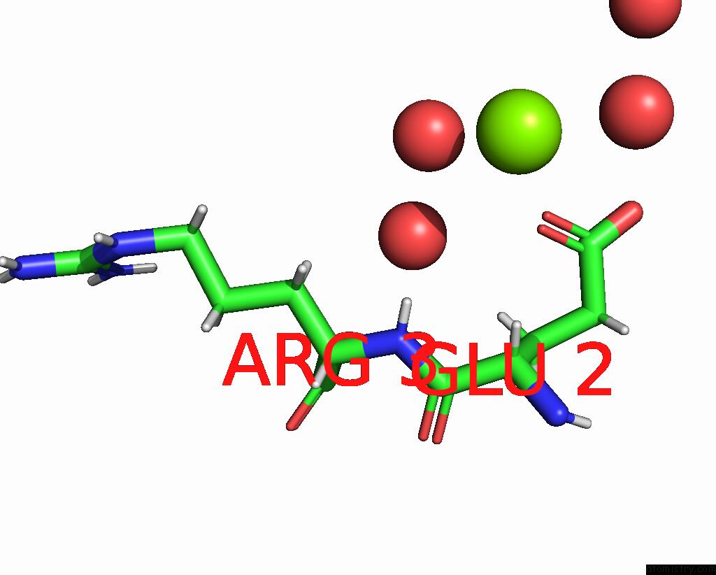
Mono view

Stereo pair view
Mono view
Stereo pair view
A full contact list of Magnesium with other atoms in the Mg binding
site number 1 of Crystal Structure of 14-3-3 Sigma in Complex with Pkr Phosphopeptide within 5.0Å range:
|
Magnesium binding site 2 out of 2 in 7obk
Go back to
Magnesium binding site 2 out
of 2 in the Crystal Structure of 14-3-3 Sigma in Complex with Pkr Phosphopeptide
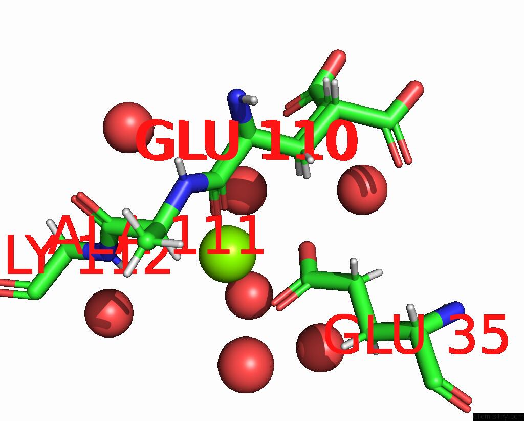
Mono view
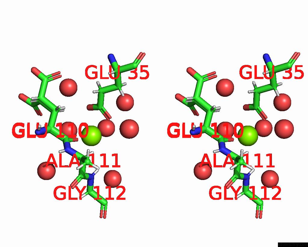
Stereo pair view
Mono view
Stereo pair view
A full contact list of Magnesium with other atoms in the Mg binding
site number 2 of Crystal Structure of 14-3-3 Sigma in Complex with Pkr Phosphopeptide within 5.0Å range:
|
Reference:
B.Andlovic,
G.Heilmann,
S.Ninck,
S.Andrei,
F.Centorrino,
Y.Higuchi,
N.Kato,
L.Brunsveld,
S.Menninger,
A.Choidas,
A.Wolf,
M.Kaiser,
J.Eickhoff,
C.Ottmann.
Inf Alpha Primes Ovarian Cancer Cells For Fusicoccin-Induced Cell Death Via Stabilization of 14-3-3 Protein-Protein Interactions To Be Published.
Page generated: Thu Oct 3 02:52:48 2024
Last articles
Mg in 2VSCMg in 2VPN
Mg in 2VRN
Mg in 2VPQ
Mg in 2VQD
Mg in 2VQ2
Mg in 2VPR
Mg in 2VPO
Mg in 2VP0
Mg in 2VOS