Magnesium »
PDB 7qoh-7qsj »
7qpr »
Magnesium in PDB 7qpr: Structure of Full Length Spot
Enzymatic activity of Structure of Full Length Spot
All present enzymatic activity of Structure of Full Length Spot:
2.7.6.5;
2.7.6.5;
Protein crystallography data
The structure of Structure of Full Length Spot, PDB code: 7qpr
was solved by
A.Garcia-Pino,
H.Tamman,
with X-Ray Crystallography technique. A brief refinement statistics is given in the table below:
| Resolution Low / High (Å) | 48.88 / 2.51 |
| Space group | P 21 21 21 |
| Cell size a, b, c (Å), α, β, γ (°) | 128.791, 133.761, 211.328, 90, 90, 90 |
| R / Rfree (%) | 22.1 / 25.8 |
Other elements in 7qpr:
The structure of Structure of Full Length Spot also contains other interesting chemical elements:
| Zinc | (Zn) | 4 atoms |
| Chlorine | (Cl) | 3 atoms |
| Manganese | (Mn) | 4 atoms |
Magnesium Binding Sites:
The binding sites of Magnesium atom in the Structure of Full Length Spot
(pdb code 7qpr). This binding sites where shown within
5.0 Angstroms radius around Magnesium atom.
In total 2 binding sites of Magnesium where determined in the Structure of Full Length Spot, PDB code: 7qpr:
Jump to Magnesium binding site number: 1; 2;
In total 2 binding sites of Magnesium where determined in the Structure of Full Length Spot, PDB code: 7qpr:
Jump to Magnesium binding site number: 1; 2;
Magnesium binding site 1 out of 2 in 7qpr
Go back to
Magnesium binding site 1 out
of 2 in the Structure of Full Length Spot

Mono view
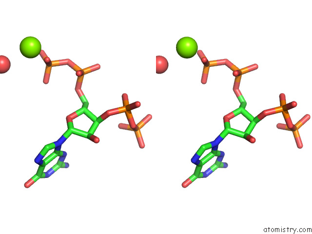
Stereo pair view
Mono view
Stereo pair view
A full contact list of Magnesium with other atoms in the Mg binding
site number 1 of Structure of Full Length Spot within 5.0Å range:
|
Magnesium binding site 2 out of 2 in 7qpr
Go back to
Magnesium binding site 2 out
of 2 in the Structure of Full Length Spot

Mono view

Stereo pair view
Mono view
Stereo pair view
A full contact list of Magnesium with other atoms in the Mg binding
site number 2 of Structure of Full Length Spot within 5.0Å range:
|
Reference:
A.Garcia-Pino,
H.Tamman.
Structure of Full Length Spot To Be Published.
Page generated: Thu Oct 3 05:55:36 2024
Last articles
I in 6QT8I in 6QFT
I in 6Q4E
I in 6Q4A
I in 6Q48
I in 6Q1L
I in 6Q3B
I in 6Q0T
I in 6Q0J
I in 6PZR