Magnesium »
PDB 7r8p-7rf0 »
7rci »
Magnesium in PDB 7rci: Crystal Structure of A PMS2 Vus with Substrate
Protein crystallography data
The structure of Crystal Structure of A PMS2 Vus with Substrate, PDB code: 7rci
was solved by
B.M.D'arcy,
A.Prakash,
with X-Ray Crystallography technique. A brief refinement statistics is given in the table below:
| Resolution Low / High (Å) | 50.47 / 2.12 |
| Space group | P 21 21 21 |
| Cell size a, b, c (Å), α, β, γ (°) | 75.059, 75.551, 135.663, 90, 90, 90 |
| R / Rfree (%) | 22.3 / 24.8 |
Magnesium Binding Sites:
The binding sites of Magnesium atom in the Crystal Structure of A PMS2 Vus with Substrate
(pdb code 7rci). This binding sites where shown within
5.0 Angstroms radius around Magnesium atom.
In total 2 binding sites of Magnesium where determined in the Crystal Structure of A PMS2 Vus with Substrate, PDB code: 7rci:
Jump to Magnesium binding site number: 1; 2;
In total 2 binding sites of Magnesium where determined in the Crystal Structure of A PMS2 Vus with Substrate, PDB code: 7rci:
Jump to Magnesium binding site number: 1; 2;
Magnesium binding site 1 out of 2 in 7rci
Go back to
Magnesium binding site 1 out
of 2 in the Crystal Structure of A PMS2 Vus with Substrate
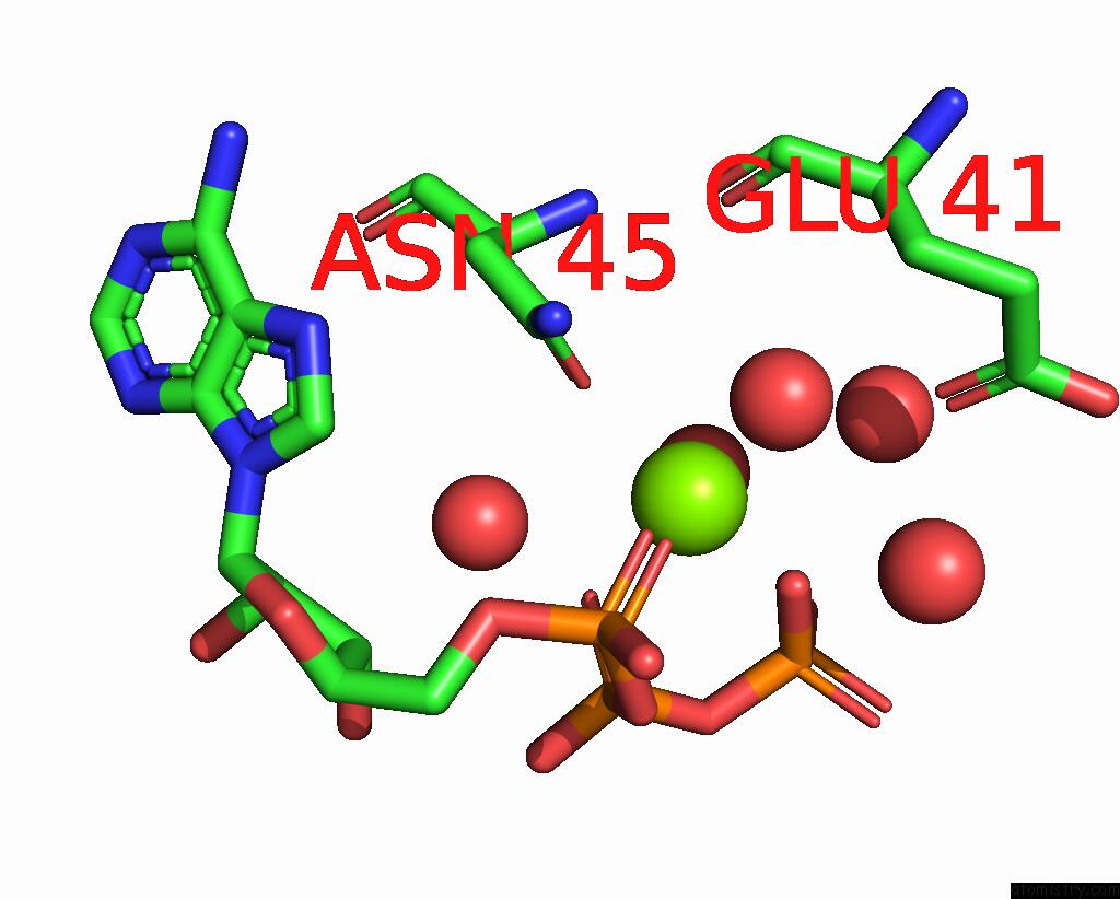
Mono view
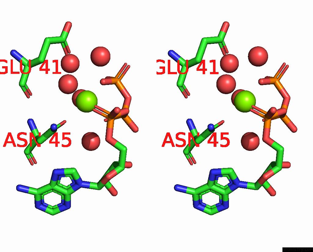
Stereo pair view
Mono view
Stereo pair view
A full contact list of Magnesium with other atoms in the Mg binding
site number 1 of Crystal Structure of A PMS2 Vus with Substrate within 5.0Å range:
|
Magnesium binding site 2 out of 2 in 7rci
Go back to
Magnesium binding site 2 out
of 2 in the Crystal Structure of A PMS2 Vus with Substrate
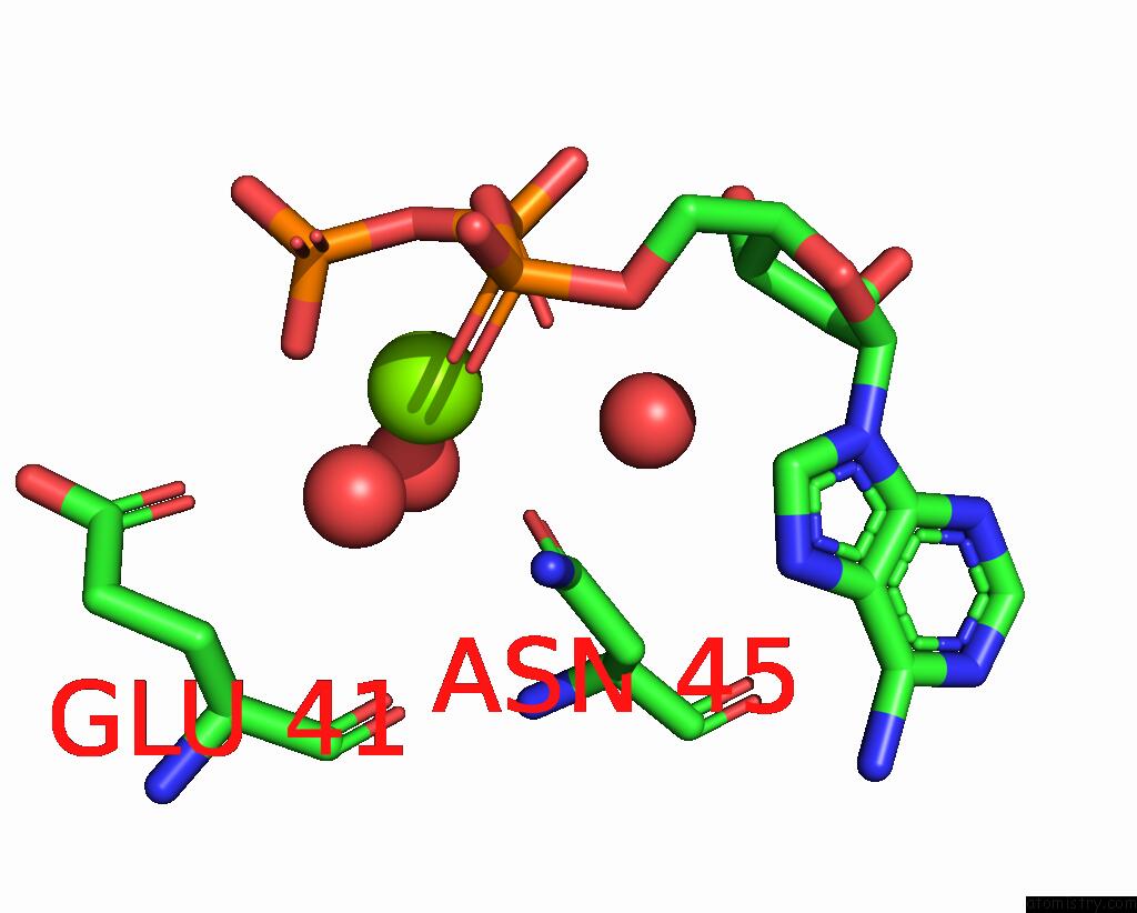
Mono view
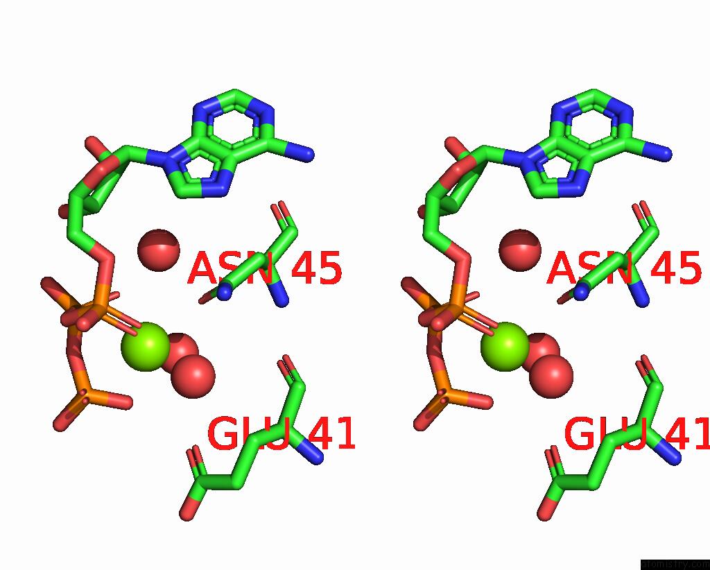
Stereo pair view
Mono view
Stereo pair view
A full contact list of Magnesium with other atoms in the Mg binding
site number 2 of Crystal Structure of A PMS2 Vus with Substrate within 5.0Å range:
|
Reference:
B.M.D'arcy,
J.Arrington,
J.Weisman,
S.B.Mcclellan,
Z.Yang,
C.Deivanayagam,
J.Blount,
A.Prakash.
PMS2 Variant Results in Loss of Atpase Activity Without Compromising Mismatch Repair. Mol Genet Genomic Med V. 10 E1908 2022.
ISSN: ISSN 2324-9269
PubMed: 35189042
DOI: 10.1002/MGG3.1908
Page generated: Thu Aug 14 14:58:25 2025
ISSN: ISSN 2324-9269
PubMed: 35189042
DOI: 10.1002/MGG3.1908
Last articles
Cl in 8QZBCl in 7HSY
Cl in 7HU9
Cl in 7HSQ
Cl in 7HRG
Cl in 7HRE
Cl in 7HRF
Cl in 7HR4
Cl in 7HQT
Cd in 9J00