Magnesium »
PDB 7tf9-7tlk »
7tih »
Magnesium in PDB 7tih: Structure of Oxidized Bovine Cytochrome C Oxidase with Reduced Metal Centers Induced By Synchrotron X-Ray Exposure
Enzymatic activity of Structure of Oxidized Bovine Cytochrome C Oxidase with Reduced Metal Centers Induced By Synchrotron X-Ray Exposure
All present enzymatic activity of Structure of Oxidized Bovine Cytochrome C Oxidase with Reduced Metal Centers Induced By Synchrotron X-Ray Exposure:
1.9.3.1;
1.9.3.1;
Protein crystallography data
The structure of Structure of Oxidized Bovine Cytochrome C Oxidase with Reduced Metal Centers Induced By Synchrotron X-Ray Exposure, PDB code: 7tih
was solved by
I.Ishigami,
D.L.Rousseau,
S.-R.Yeh,
S.Russi,
A.Cohen,
with X-Ray Crystallography technique. A brief refinement statistics is given in the table below:
| Resolution Low / High (Å) | 39.34 / 2.35 |
| Space group | P 21 21 21 |
| Cell size a, b, c (Å), α, β, γ (°) | 178.037, 182.696, 205.854, 90, 90, 90 |
| R / Rfree (%) | 19.3 / 23.5 |
Other elements in 7tih:
The structure of Structure of Oxidized Bovine Cytochrome C Oxidase with Reduced Metal Centers Induced By Synchrotron X-Ray Exposure also contains other interesting chemical elements:
| Iron | (Fe) | 4 atoms |
| Copper | (Cu) | 6 atoms |
| Zinc | (Zn) | 2 atoms |
| Sodium | (Na) | 2 atoms |
Magnesium Binding Sites:
The binding sites of Magnesium atom in the Structure of Oxidized Bovine Cytochrome C Oxidase with Reduced Metal Centers Induced By Synchrotron X-Ray Exposure
(pdb code 7tih). This binding sites where shown within
5.0 Angstroms radius around Magnesium atom.
In total 2 binding sites of Magnesium where determined in the Structure of Oxidized Bovine Cytochrome C Oxidase with Reduced Metal Centers Induced By Synchrotron X-Ray Exposure, PDB code: 7tih:
Jump to Magnesium binding site number: 1; 2;
In total 2 binding sites of Magnesium where determined in the Structure of Oxidized Bovine Cytochrome C Oxidase with Reduced Metal Centers Induced By Synchrotron X-Ray Exposure, PDB code: 7tih:
Jump to Magnesium binding site number: 1; 2;
Magnesium binding site 1 out of 2 in 7tih
Go back to
Magnesium binding site 1 out
of 2 in the Structure of Oxidized Bovine Cytochrome C Oxidase with Reduced Metal Centers Induced By Synchrotron X-Ray Exposure
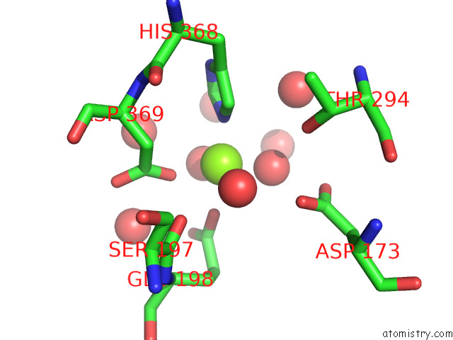
Mono view
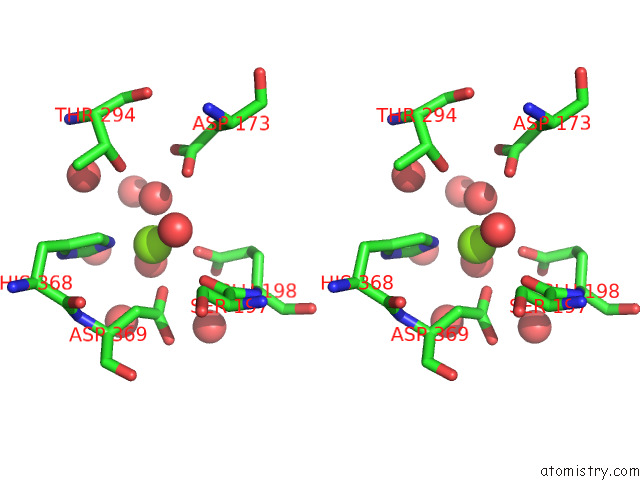
Stereo pair view
Mono view
Stereo pair view
A full contact list of Magnesium with other atoms in the Mg binding
site number 1 of Structure of Oxidized Bovine Cytochrome C Oxidase with Reduced Metal Centers Induced By Synchrotron X-Ray Exposure within 5.0Å range:
|
Magnesium binding site 2 out of 2 in 7tih
Go back to
Magnesium binding site 2 out
of 2 in the Structure of Oxidized Bovine Cytochrome C Oxidase with Reduced Metal Centers Induced By Synchrotron X-Ray Exposure
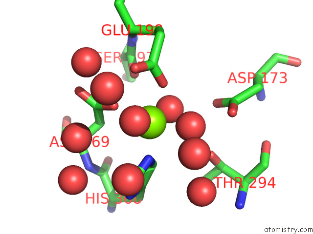
Mono view
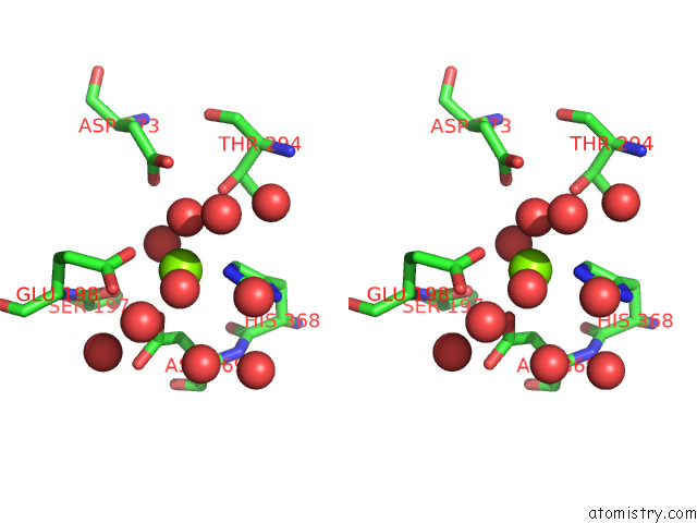
Stereo pair view
Mono view
Stereo pair view
A full contact list of Magnesium with other atoms in the Mg binding
site number 2 of Structure of Oxidized Bovine Cytochrome C Oxidase with Reduced Metal Centers Induced By Synchrotron X-Ray Exposure within 5.0Å range:
|
Reference:
I.Ishigami,
S.Russi,
A.Cohen,
S.R.Yeh,
D.L.Rousseau.
Temperature-Dependent Structural Transition Following X-Ray-Induced Metal Center Reduction in Oxidized Cytochrome C Oxidase. J.Biol.Chem. V. 298 01799 2022.
ISSN: ESSN 1083-351X
PubMed: 35257742
DOI: 10.1016/J.JBC.2022.101799
Page generated: Thu Oct 3 09:17:51 2024
ISSN: ESSN 1083-351X
PubMed: 35257742
DOI: 10.1016/J.JBC.2022.101799
Last articles
I in 3S6LI in 3T96
I in 3SV5
I in 3T3W
I in 3T3H
I in 3SIA
I in 3SSL
I in 3SSY
I in 3SEX
I in 3SLS