Magnesium »
PDB 7vx8-7w31 »
7w19 »
Magnesium in PDB 7w19: Crystal Structure of Acinetobacter Baumannii Mph-E
Protein crystallography data
The structure of Crystal Structure of Acinetobacter Baumannii Mph-E, PDB code: 7w19
was solved by
Q.Qi,
L.Kuang,
Y.Jiang,
with X-Ray Crystallography technique. A brief refinement statistics is given in the table below:
| Resolution Low / High (Å) | 46.11 / 1.90 |
| Space group | C 2 2 21 |
| Cell size a, b, c (Å), α, β, γ (°) | 86.844, 142.172, 92.229, 90, 90, 90 |
| R / Rfree (%) | 22.4 / 26.5 |
Other elements in 7w19:
The structure of Crystal Structure of Acinetobacter Baumannii Mph-E also contains other interesting chemical elements:
| Calcium | (Ca) | 2 atoms |
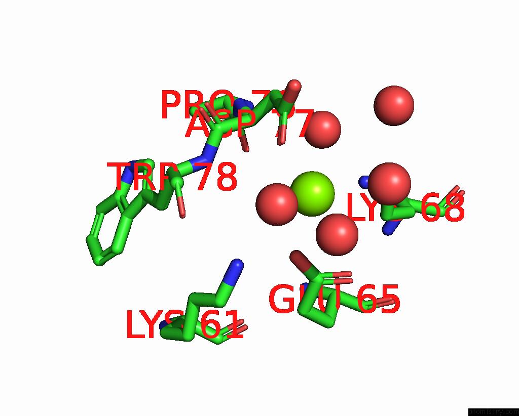
Magnesium Binding Sites:
The binding sites of Magnesium atom in the Crystal Structure of Acinetobacter Baumannii Mph-E
(pdb code 7w19). This binding sites where shown within
5.0 Angstroms radius around Magnesium atom.
In total only one binding site of Magnesium was determined in the Crystal Structure of Acinetobacter Baumannii Mph-E, PDB code: 7w19:
In total only one binding site of Magnesium was determined in the Crystal Structure of Acinetobacter Baumannii Mph-E, PDB code: 7w19:
Magnesium binding site 1 out of 1 in 7w19
Go back to
Magnesium binding site 1 out
of 1 in the Crystal Structure of Acinetobacter Baumannii Mph-E
Mono view

Stereo pair view
Mono view
Stereo pair view
A full contact list of Magnesium with other atoms in the Mg binding
site number 1 of Crystal Structure of Acinetobacter Baumannii Mph-E within 5.0Å range:
|
Reference:
Q.Qi,
L.Kuang,
Y.Jiang.
Crystal Structure of the Acinetobacter Baumannii Macrolide Phosphotransferases E Reveal the Novel Catalysis Mechanism To Be Published.
Page generated: Thu Oct 3 11:01:07 2024
Last articles
Mg in 4V1MMg in 4V0R
Mg in 4V0O
Mg in 4V0N
Mg in 4V0M
Mg in 4V0L
Mg in 4V08
Mg in 4V03
Mg in 4V07
Mg in 4V02