Magnesium »
PDB 7wrx-7x75 »
7wyg »
Magnesium in PDB 7wyg: Crystal Structure of P450BSBETA-L78I/Q85H/G290I Variant in Complex with Palmitic Acid.
Protein crystallography data
The structure of Crystal Structure of P450BSBETA-L78I/Q85H/G290I Variant in Complex with Palmitic Acid., PDB code: 7wyg
was solved by
F.Li,
C.He,
X.Wang,
with X-Ray Crystallography technique. A brief refinement statistics is given in the table below:
| Resolution Low / High (Å) | 50.98 / 2.00 |
| Space group | P 3 2 1 |
| Cell size a, b, c (Å), α, β, γ (°) | 125.646, 125.646, 145.891, 90, 90, 120 |
| R / Rfree (%) | 17.4 / 20.8 |
Other elements in 7wyg:
The structure of Crystal Structure of P450BSBETA-L78I/Q85H/G290I Variant in Complex with Palmitic Acid. also contains other interesting chemical elements:
| Iron | (Fe) | 2 atoms |
Magnesium Binding Sites:
The binding sites of Magnesium atom in the Crystal Structure of P450BSBETA-L78I/Q85H/G290I Variant in Complex with Palmitic Acid.
(pdb code 7wyg). This binding sites where shown within
5.0 Angstroms radius around Magnesium atom.
In total 2 binding sites of Magnesium where determined in the Crystal Structure of P450BSBETA-L78I/Q85H/G290I Variant in Complex with Palmitic Acid., PDB code: 7wyg:
Jump to Magnesium binding site number: 1; 2;
In total 2 binding sites of Magnesium where determined in the Crystal Structure of P450BSBETA-L78I/Q85H/G290I Variant in Complex with Palmitic Acid., PDB code: 7wyg:
Jump to Magnesium binding site number: 1; 2;
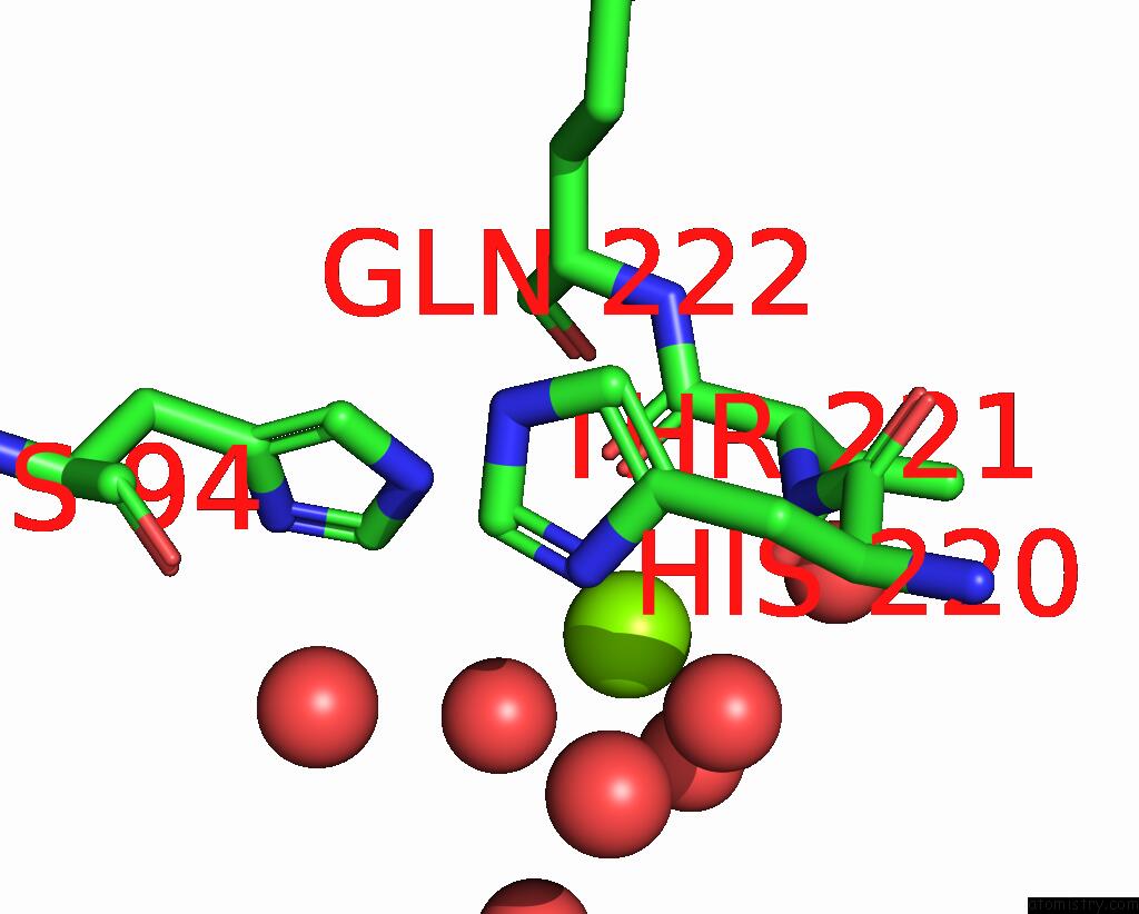
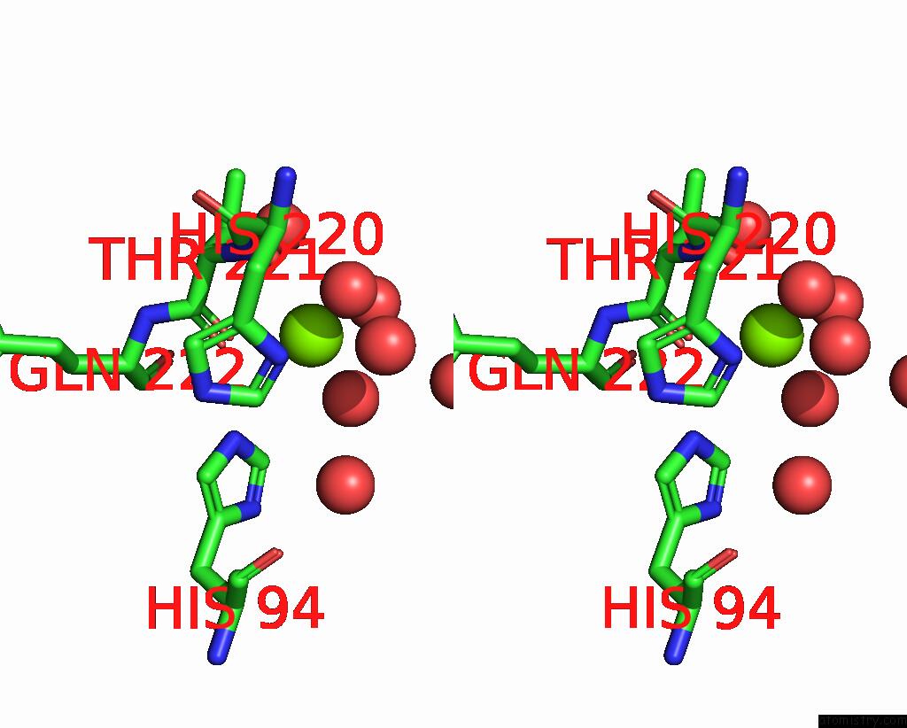
Magnesium binding site 1 out of 2 in 7wyg
Go back to
Magnesium binding site 1 out
of 2 in the Crystal Structure of P450BSBETA-L78I/Q85H/G290I Variant in Complex with Palmitic Acid.

Mono view

Stereo pair view
Mono view
Stereo pair view
A full contact list of Magnesium with other atoms in the Mg binding
site number 1 of Crystal Structure of P450BSBETA-L78I/Q85H/G290I Variant in Complex with Palmitic Acid. within 5.0Å range:
|
Magnesium binding site 2 out of 2 in 7wyg
Go back to
Magnesium binding site 2 out
of 2 in the Crystal Structure of P450BSBETA-L78I/Q85H/G290I Variant in Complex with Palmitic Acid.
Mono view
Stereo pair view
Mono view
Stereo pair view
A full contact list of Magnesium with other atoms in the Mg binding
site number 2 of Crystal Structure of P450BSBETA-L78I/Q85H/G290I Variant in Complex with Palmitic Acid. within 5.0Å range:
|
Reference:
K.Zhang,
A.Yu,
X.Chu,
F.Li,
J.Liu,
L.Liu,
W.J.Bai,
C.He,
X.Wang.
Biocatalytic Enantioselective Beta-Hydroxylation of Unactivated C-H Bonds in Aliphatic Carboxylic Acids. Angew.Chem.Int.Ed.Engl. V. 61 04290 2022.
ISSN: ESSN 1521-3773
PubMed: 35536725
DOI: 10.1002/ANIE.202204290
Page generated: Thu Aug 14 17:41:10 2025
ISSN: ESSN 1521-3773
PubMed: 35536725
DOI: 10.1002/ANIE.202204290
Last articles
Xe in 1C6EXe in 1C68
Xe in 1C6B
Xe in 1C10
Xe in 1C65
Xe in 1C62
Xe in 1C3L
Xe in 1C1M
W in 8PRO
W in 9FPP