Magnesium »
PDB 8a65-8ac2 »
8a9n »
Magnesium in PDB 8a9n: Structure of Dpa Polyamine Acetyltransferase in Complex with 1,3-Dap
Protein crystallography data
The structure of Structure of Dpa Polyamine Acetyltransferase in Complex with 1,3-Dap, PDB code: 8a9n
was solved by
A.Garcia-Pino,
D.Jurenas,
with X-Ray Crystallography technique. A brief refinement statistics is given in the table below:
| Resolution Low / High (Å) | 41.66 / 1.85 |
| Space group | P 1 21 1 |
| Cell size a, b, c (Å), α, β, γ (°) | 42.87, 68.08, 55.74, 90, 109.12, 90 |
| R / Rfree (%) | 20.6 / 25.1 |
Magnesium Binding Sites:
The binding sites of Magnesium atom in the Structure of Dpa Polyamine Acetyltransferase in Complex with 1,3-Dap
(pdb code 8a9n). This binding sites where shown within
5.0 Angstroms radius around Magnesium atom.
In total 2 binding sites of Magnesium where determined in the Structure of Dpa Polyamine Acetyltransferase in Complex with 1,3-Dap, PDB code: 8a9n:
Jump to Magnesium binding site number: 1; 2;
In total 2 binding sites of Magnesium where determined in the Structure of Dpa Polyamine Acetyltransferase in Complex with 1,3-Dap, PDB code: 8a9n:
Jump to Magnesium binding site number: 1; 2;
Magnesium binding site 1 out of 2 in 8a9n
Go back to
Magnesium binding site 1 out
of 2 in the Structure of Dpa Polyamine Acetyltransferase in Complex with 1,3-Dap
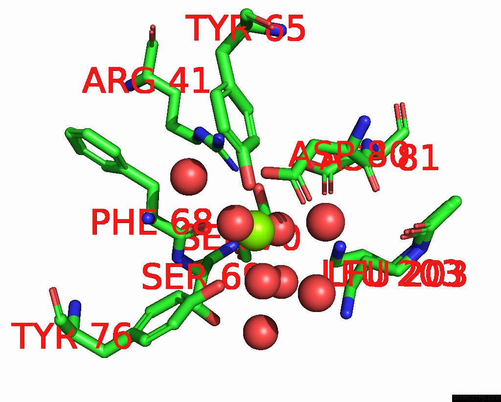
Mono view
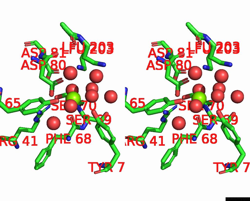
Stereo pair view
Mono view
Stereo pair view
A full contact list of Magnesium with other atoms in the Mg binding
site number 1 of Structure of Dpa Polyamine Acetyltransferase in Complex with 1,3-Dap within 5.0Å range:
|
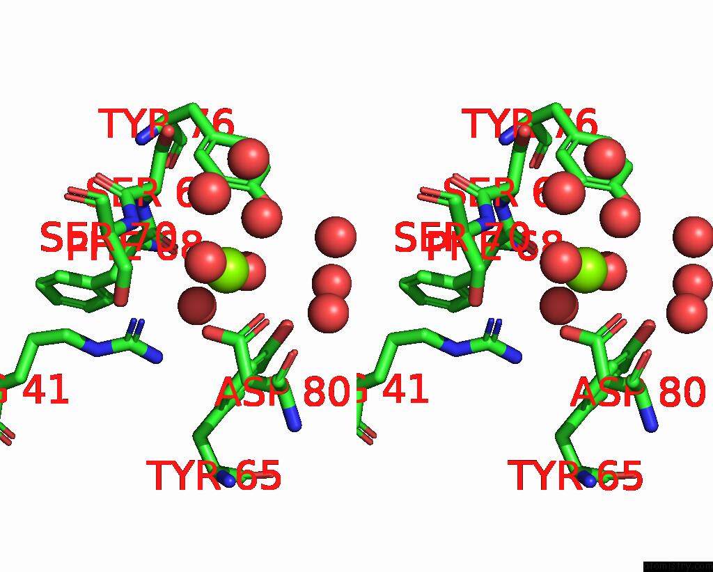
Magnesium binding site 2 out of 2 in 8a9n
Go back to
Magnesium binding site 2 out
of 2 in the Structure of Dpa Polyamine Acetyltransferase in Complex with 1,3-Dap
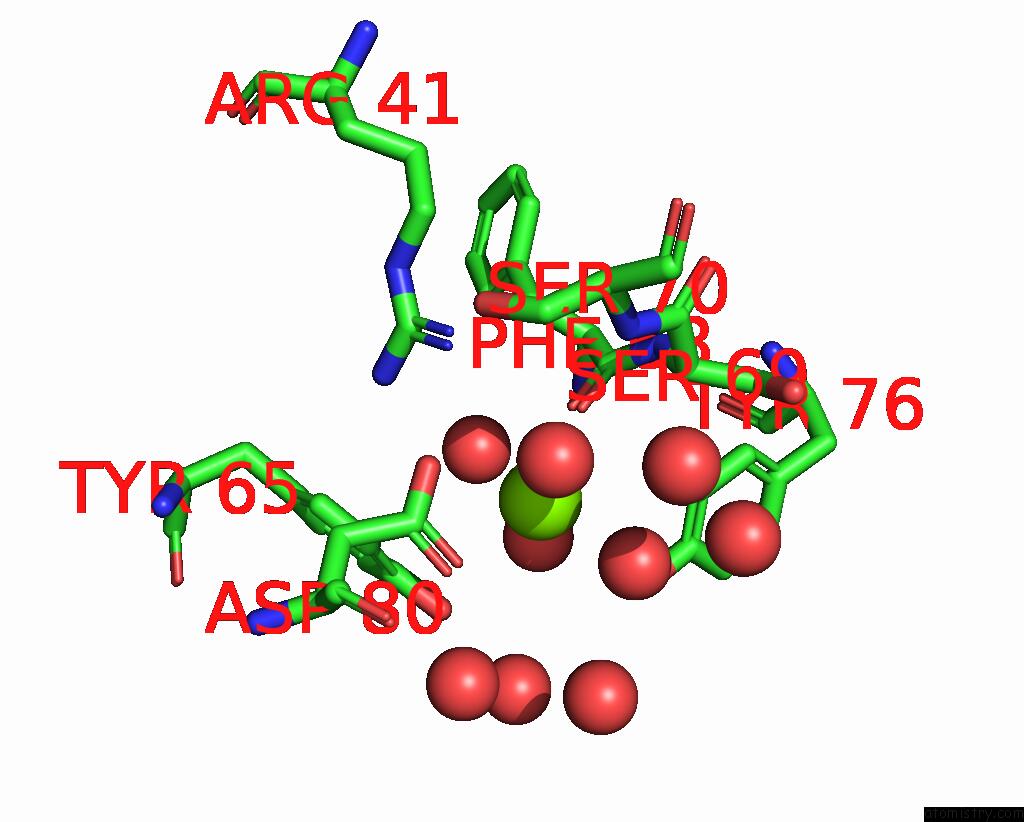
Mono view
Stereo pair view
Mono view
Stereo pair view
A full contact list of Magnesium with other atoms in the Mg binding
site number 2 of Structure of Dpa Polyamine Acetyltransferase in Complex with 1,3-Dap within 5.0Å range:
|
Reference:
A.Garcia-Pino,
D.Jurenas.
Structure of Dpa Polyamine Acetyltransferase in Complex with 1,3-Dap To Be Published.
Page generated: Thu Oct 3 18:00:12 2024
Last articles
Mg in 5SEQMg in 5SEO
Mg in 5SEP
Mg in 5SEN
Mg in 5SEM
Mg in 5SEL
Mg in 5SEK
Mg in 5SEJ
Mg in 5SEI
Mg in 5SEH