Magnesium »
PDB 8c7c-8cgk »
8cga »
Magnesium in PDB 8cga: Structure of Mycobacterium Tuberculosis Dutpase Delta 133A-137S Mutant
Enzymatic activity of Structure of Mycobacterium Tuberculosis Dutpase Delta 133A-137S Mutant
All present enzymatic activity of Structure of Mycobacterium Tuberculosis Dutpase Delta 133A-137S Mutant:
3.6.1.23;
3.6.1.23;
Protein crystallography data
The structure of Structure of Mycobacterium Tuberculosis Dutpase Delta 133A-137S Mutant, PDB code: 8cga
was solved by
Z.S.Toth,
A.Benedek,
I.Leveles,
B.G.Vertessy,
with X-Ray Crystallography technique. A brief refinement statistics is given in the table below:
| Resolution Low / High (Å) | 47.85 / 1.30 |
| Space group | P 63 |
| Cell size a, b, c (Å), α, β, γ (°) | 55.249, 55.249, 83.751, 90, 90, 120 |
| R / Rfree (%) | 14.1 / 17 |
Magnesium Binding Sites:
The binding sites of Magnesium atom in the Structure of Mycobacterium Tuberculosis Dutpase Delta 133A-137S Mutant
(pdb code 8cga). This binding sites where shown within
5.0 Angstroms radius around Magnesium atom.
In total only one binding site of Magnesium was determined in the Structure of Mycobacterium Tuberculosis Dutpase Delta 133A-137S Mutant, PDB code: 8cga:
In total only one binding site of Magnesium was determined in the Structure of Mycobacterium Tuberculosis Dutpase Delta 133A-137S Mutant, PDB code: 8cga:
Magnesium binding site 1 out of 1 in 8cga
Go back to
Magnesium binding site 1 out
of 1 in the Structure of Mycobacterium Tuberculosis Dutpase Delta 133A-137S Mutant
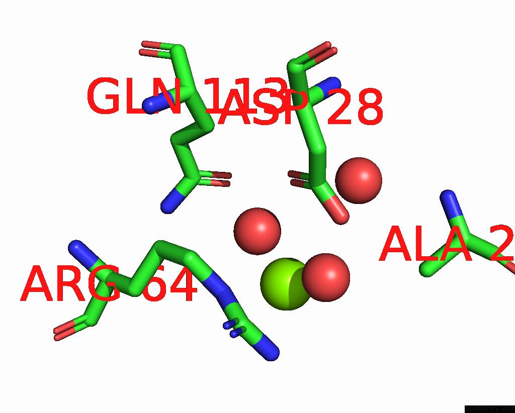
Mono view
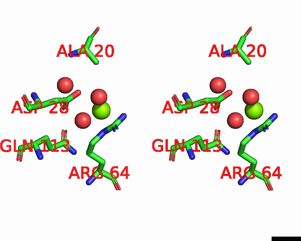
Stereo pair view
Mono view
Stereo pair view
A full contact list of Magnesium with other atoms in the Mg binding
site number 1 of Structure of Mycobacterium Tuberculosis Dutpase Delta 133A-137S Mutant within 5.0Å range:
|
Reference:
Z.S.Toth,
A.Benedek,
I.Leveles,
B.G.Vertessy.
Structure of Mycobacterium Tuberculosis Dutpase Delta 133A-137S Mutant To Be Published.
Page generated: Thu Oct 3 20:48:17 2024
Last articles
Mg in 4NDRMg in 4NDN
Mg in 4NDQ
Mg in 4NC4
Mg in 4NDP
Mg in 4NDO
Mg in 4NCN
Mg in 4NCJ
Mg in 4NCL
Mg in 4NCK