Magnesium »
PDB 8gi3-8gwe »
8gju »
Magnesium in PDB 8gju: Crystal Structure of Human Methylmalonyl-Coa Mutase (Mmut) in Complex with Methylmalonic Acidemia Type A Protein (Mmaa), Coenzyme A, and Gdp
Enzymatic activity of Crystal Structure of Human Methylmalonyl-Coa Mutase (Mmut) in Complex with Methylmalonic Acidemia Type A Protein (Mmaa), Coenzyme A, and Gdp
All present enzymatic activity of Crystal Structure of Human Methylmalonyl-Coa Mutase (Mmut) in Complex with Methylmalonic Acidemia Type A Protein (Mmaa), Coenzyme A, and Gdp:
5.4.99.2;
5.4.99.2;
Protein crystallography data
The structure of Crystal Structure of Human Methylmalonyl-Coa Mutase (Mmut) in Complex with Methylmalonic Acidemia Type A Protein (Mmaa), Coenzyme A, and Gdp, PDB code: 8gju
was solved by
R.M.Mascarenhas,
M.Ruetz,
H.Gouda,
M.Yaw,
R.Banerjee,
with X-Ray Crystallography technique. A brief refinement statistics is given in the table below:
| Resolution Low / High (Å) | 80.62 / 2.79 |
| Space group | P 1 21 1 |
| Cell size a, b, c (Å), α, β, γ (°) | 95.806, 221.872, 121.824, 90, 105.53, 90 |
| R / Rfree (%) | 22.9 / 27.6 |
Magnesium Binding Sites:
The binding sites of Magnesium atom in the Crystal Structure of Human Methylmalonyl-Coa Mutase (Mmut) in Complex with Methylmalonic Acidemia Type A Protein (Mmaa), Coenzyme A, and Gdp
(pdb code 8gju). This binding sites where shown within
5.0 Angstroms radius around Magnesium atom.
In total 4 binding sites of Magnesium where determined in the Crystal Structure of Human Methylmalonyl-Coa Mutase (Mmut) in Complex with Methylmalonic Acidemia Type A Protein (Mmaa), Coenzyme A, and Gdp, PDB code: 8gju:
Jump to Magnesium binding site number: 1; 2; 3; 4;
In total 4 binding sites of Magnesium where determined in the Crystal Structure of Human Methylmalonyl-Coa Mutase (Mmut) in Complex with Methylmalonic Acidemia Type A Protein (Mmaa), Coenzyme A, and Gdp, PDB code: 8gju:
Jump to Magnesium binding site number: 1; 2; 3; 4;
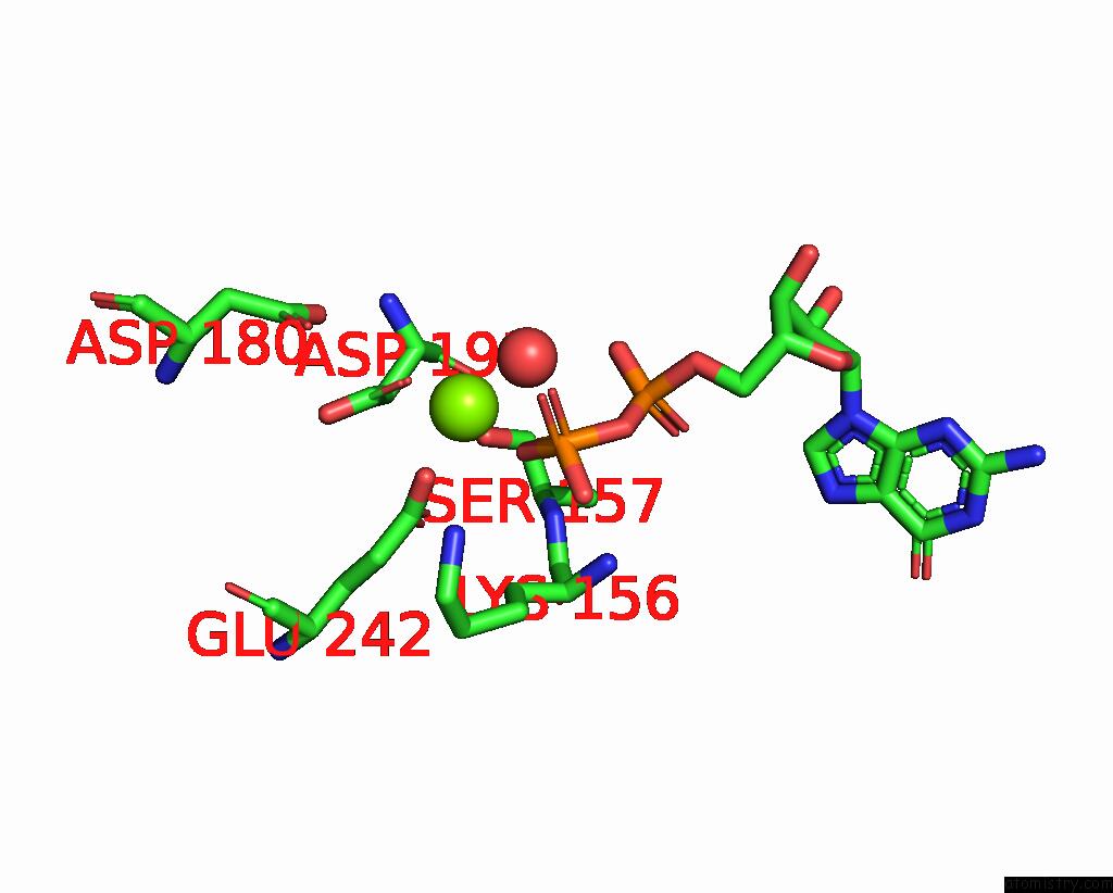
Magnesium binding site 1 out of 4 in 8gju
Go back to
Magnesium binding site 1 out
of 4 in the Crystal Structure of Human Methylmalonyl-Coa Mutase (Mmut) in Complex with Methylmalonic Acidemia Type A Protein (Mmaa), Coenzyme A, and Gdp

Mono view

Stereo pair view
Mono view
Stereo pair view
A full contact list of Magnesium with other atoms in the Mg binding
site number 1 of Crystal Structure of Human Methylmalonyl-Coa Mutase (Mmut) in Complex with Methylmalonic Acidemia Type A Protein (Mmaa), Coenzyme A, and Gdp within 5.0Å range:
|
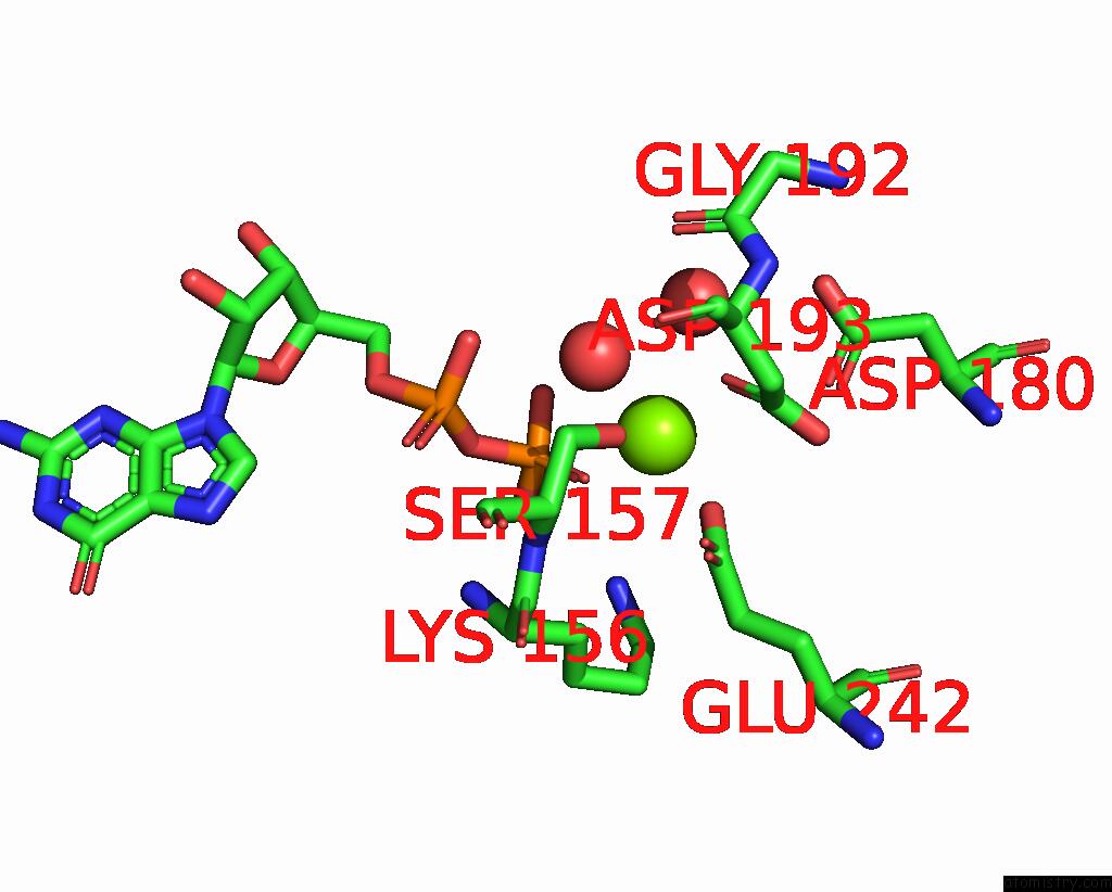
Magnesium binding site 2 out of 4 in 8gju
Go back to
Magnesium binding site 2 out
of 4 in the Crystal Structure of Human Methylmalonyl-Coa Mutase (Mmut) in Complex with Methylmalonic Acidemia Type A Protein (Mmaa), Coenzyme A, and Gdp
Mono view

Stereo pair view
Mono view
Stereo pair view
A full contact list of Magnesium with other atoms in the Mg binding
site number 2 of Crystal Structure of Human Methylmalonyl-Coa Mutase (Mmut) in Complex with Methylmalonic Acidemia Type A Protein (Mmaa), Coenzyme A, and Gdp within 5.0Å range:
|
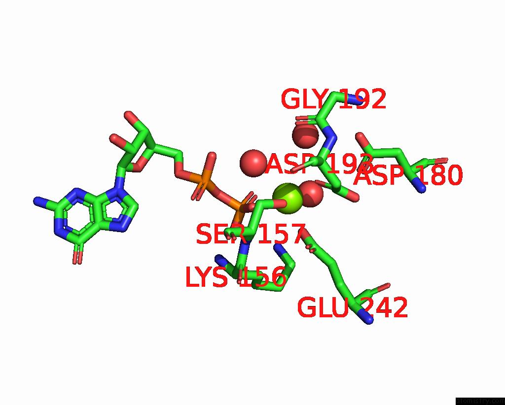
Magnesium binding site 3 out of 4 in 8gju
Go back to
Magnesium binding site 3 out
of 4 in the Crystal Structure of Human Methylmalonyl-Coa Mutase (Mmut) in Complex with Methylmalonic Acidemia Type A Protein (Mmaa), Coenzyme A, and Gdp
Mono view

Stereo pair view
Mono view
Stereo pair view
A full contact list of Magnesium with other atoms in the Mg binding
site number 3 of Crystal Structure of Human Methylmalonyl-Coa Mutase (Mmut) in Complex with Methylmalonic Acidemia Type A Protein (Mmaa), Coenzyme A, and Gdp within 5.0Å range:
|
Magnesium binding site 4 out of 4 in 8gju
Go back to
Magnesium binding site 4 out
of 4 in the Crystal Structure of Human Methylmalonyl-Coa Mutase (Mmut) in Complex with Methylmalonic Acidemia Type A Protein (Mmaa), Coenzyme A, and Gdp
Mono view
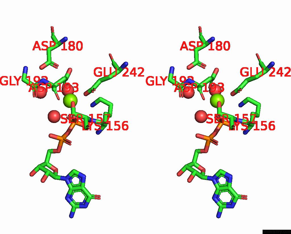
Stereo pair view
Mono view
Stereo pair view
A full contact list of Magnesium with other atoms in the Mg binding
site number 4 of Crystal Structure of Human Methylmalonyl-Coa Mutase (Mmut) in Complex with Methylmalonic Acidemia Type A Protein (Mmaa), Coenzyme A, and Gdp within 5.0Å range:
|
Reference:
R.Mascarenhas,
M.Ruetz,
H.Gouda,
N.Heitman,
M.Yaw,
R.Banerjee.
Architecture of the Human G-Protein-Methylmalonyl-Coa Mutase Nanoassembly For B 12 Delivery and Repair. Nat Commun V. 14 4332 2023.
ISSN: ESSN 2041-1723
PubMed: 37468522
DOI: 10.1038/S41467-023-40077-4
Page generated: Fri Oct 4 03:51:20 2024
ISSN: ESSN 2041-1723
PubMed: 37468522
DOI: 10.1038/S41467-023-40077-4
Last articles
Mg in 7DKZMg in 7DDQ
Mg in 7DIY
Mg in 7DID
Mg in 7DHW
Mg in 7DIC
Mg in 7DFU
Mg in 7DFJ
Mg in 7DFK
Mg in 7DFH