Magnesium »
PDB 8h6s-8hha »
8h6s »
Magnesium in PDB 8h6s: Structure of Acyltransferase Vink in Complex with the Loading Acyl Carrier Protein of Vicenistatin Pks
Protein crystallography data
The structure of Structure of Acyltransferase Vink in Complex with the Loading Acyl Carrier Protein of Vicenistatin Pks, PDB code: 8h6s
was solved by
K.Kawada,
A.Miyanaga,
T.Chisuga,
F.Kudo,
T.Eguchi,
with X-Ray Crystallography technique. A brief refinement statistics is given in the table below:
| Resolution Low / High (Å) | 46.01 / 3.00 |
| Space group | P 21 21 21 |
| Cell size a, b, c (Å), α, β, γ (°) | 78.363, 90.626, 113.511, 90, 90, 90 |
| R / Rfree (%) | 21 / 26.8 |
Magnesium Binding Sites:
The binding sites of Magnesium atom in the Structure of Acyltransferase Vink in Complex with the Loading Acyl Carrier Protein of Vicenistatin Pks
(pdb code 8h6s). This binding sites where shown within
5.0 Angstroms radius around Magnesium atom.
In total 2 binding sites of Magnesium where determined in the Structure of Acyltransferase Vink in Complex with the Loading Acyl Carrier Protein of Vicenistatin Pks, PDB code: 8h6s:
Jump to Magnesium binding site number: 1; 2;
In total 2 binding sites of Magnesium where determined in the Structure of Acyltransferase Vink in Complex with the Loading Acyl Carrier Protein of Vicenistatin Pks, PDB code: 8h6s:
Jump to Magnesium binding site number: 1; 2;
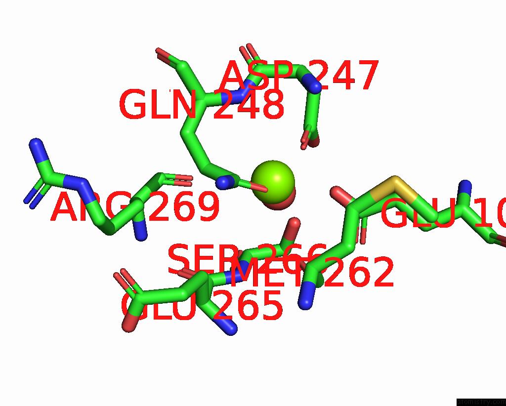
Magnesium binding site 1 out of 2 in 8h6s
Go back to
Magnesium binding site 1 out
of 2 in the Structure of Acyltransferase Vink in Complex with the Loading Acyl Carrier Protein of Vicenistatin Pks

Mono view

Stereo pair view
Mono view
Stereo pair view
A full contact list of Magnesium with other atoms in the Mg binding
site number 1 of Structure of Acyltransferase Vink in Complex with the Loading Acyl Carrier Protein of Vicenistatin Pks within 5.0Å range:
|
Magnesium binding site 2 out of 2 in 8h6s
Go back to
Magnesium binding site 2 out
of 2 in the Structure of Acyltransferase Vink in Complex with the Loading Acyl Carrier Protein of Vicenistatin Pks
Mono view

Stereo pair view
Mono view
Stereo pair view
A full contact list of Magnesium with other atoms in the Mg binding
site number 2 of Structure of Acyltransferase Vink in Complex with the Loading Acyl Carrier Protein of Vicenistatin Pks within 5.0Å range:
|
Reference:
A.Miyanaga,
K.Kawada,
T.Chisuga,
F.Kudo,
T.Eguchi.
Structural Basis of Transient Interactions of Acyltransferase Vink with the Loading Acyl Carrier Protein of the Vicenistatin Modular Polyketide Synthase. Biochemistry 2022.
ISSN: ISSN 0006-2960
PubMed: 36512613
DOI: 10.1021/ACS.BIOCHEM.2C00645
Page generated: Fri Aug 15 06:18:06 2025
ISSN: ISSN 0006-2960
PubMed: 36512613
DOI: 10.1021/ACS.BIOCHEM.2C00645
Last articles
Mg in 8IWXMg in 8IUN
Mg in 8IUG
Mg in 8IWU
Mg in 8IWT
Mg in 8IWS
Mg in 8IWR
Mg in 8IWN
Mg in 8IU7
Mg in 8IUH